A 5-week-old girl, who appeared to be healthy at birth, develops diarrhea and vomiting a few days after birth. Your current examination reveals that she has hepatomegaly, jaundice, and early cataract formation and is not meeting developmental milestones. You suspect that she has which of the following conditions?
A. galactosemia
B. Hurler syndrome
C. pyloric stenosis
D. Tay-Sachs disease
E. type I glycogenosis
Correct Answer: A
Section: Pathology and Path physiology Galactosemia is an autosomal recessive disorder due (in this more common and more severe form of the disease) to a lack of galactose- 1-phosphate uridyl transferase. This results in the formation and accumulation of galactose metabolites. If the infant's diet is not modified to exclude milk products, this will result in damage to the liver (fatty change, cholestasis, cirrhosis, liver failure), eyes (cataract formation), and brain (mental retardation). Hurler syndrome (choice B) is a severe form of mucopolysaccharidosis that typically becomes apparent between 6 months and 2 years of age. Prominent features include coarse facies, dwarfism, organomegaly, cataracts, and mental retardation, not diarrhea, vomiting, and jaundice. Pyloric stenosis (choice C) can occur as a congenital condition, more frequently in baby boys (M:F = 4:1). It is marked by projectile vomiting in the first month of life, but not the other findings in this case. Tay-Sachs disease (choice D) is a lipid storage disease due to a deficiency of hexosaminidase A. There is an inexorable deterioration of mental and motor functions within a few months of birth culminating in a vegetative state and death within 3 or 4 years. Type I glycogen storage disease or von Gierke disease (choice E) is due to a deficiency of glucose-6- phosphatase and usually becomes apparent in the first year of life as hypoglycemia and/or hepatomegaly.
Question 302:
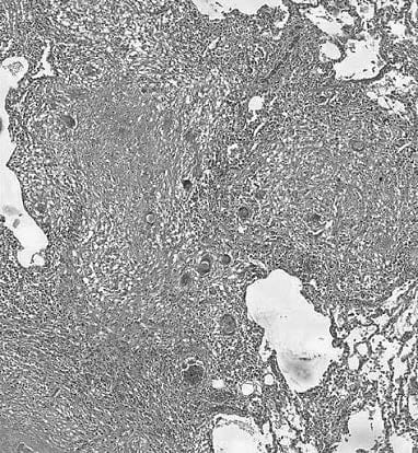
A 47-year-old man has been chronically ill for the past 18 months. He undergoes a lung biopsy and a ection is shown in below figure. Which of the following is the most likely diagnosis?
A. aspergillosis
B. leprosy
C. pneumocystosis
D. sarcoidosis
E. tuberculosis
Correct Answer: E
Section: Pathology and Path physiology Figure shows a microscopic section of lung in which there are a number of necrotizing granulomascontaining giant cells. Aspergillosis (choice A) and pneumocystosis (choice C) are not associated with granuloma formation. Leprosy (choice B) does (in tuberculoid leprosy) form granulomas and may be found in the upper airways, but does not involve viscera such as the lung. Sarcoidosis (choice D) is a granulomatous disease of unknown etiology that can involve the lung and many other organs but the granulomas are non-necrotizing.
Question 303:
A 32-year-old man presents with diarrhea and symptoms of peptic ulcer disease. Endoscopy reveals two ulcers, one in the first portion of the duodenum and one in the midduodenum. However, they do not respond to the usual peptic ulcer treatment programs. The most likely explanation for the findings in this patient is which of the following?
A. antibodies to intrinsic factor
B. ectopic hypersecretion of gastrin
C. gastric mucosal atrophy
D. pressure ulceration from bezoars
E. vascular abnormality
Correct Answer: B
Section: Pathology and Path physiology Peptic ulcerations seen in ZollingerEllison syndrome are due to ectopic hypersecretion of gastrin. An islet cell tumor of the pancreas is the most frequent ectopic site. Antibodies to intrinsic factor (choice A) are seen with pernicious anemia and usually cause gastric mucosal atrophy and metaplasia. Gastric mucosal hypertrophy, not atrophy (choice C), is the expected result with an increased secretion of gastrin as is seen with ZollingerEllison syndrome. Pressure ulceration from bezoars (choice D) and vascular abnormalities (choice E) are not the etiology of the peptic ulcerations seen with Zollinger Ellison syndrome.
Question 304:
A 76-year-old woman suffers a massive myocardial infarct and dies in cardiogenic shock 20 hours after its onset. Microscopic examination of her infarcted myocardium would be expected to demonstrate which of the following?
A. abundant neutrophils and monocytes
B. coagulative necrosis with few neutrophils
C. fibrosis and collagen deposition
D. monocytes and neovascularization
E. plasma cells and caseous necrosis
Correct Answer: B
Section: Pathology and Path physiology A 20-hour-old ischemic infarct of the myocardium should demonstrate coagulative necrosis without much of an inflammatory response. Abundant neutrophils and monocytes (choice A) typically are seen about 24 days after an infarction. Fibrosis and collagen deposition (choice C) are late healing phenomena that do not begin until at least 1 week after the infarct has occurred. Monocytic infiltration and neovascularization (choice D) usually occur about 36 days after an infarction. Plasma cells and caseous necrosis (choice E) are not seen with ischemic myocardial damage. Plasma cells are typically seen in areas of chronic inflammation and caseous necrosis is found in granulomas produced in response to tuberculosis and certain fungal infections.
Question 305:
A 32-year-old male cyclist is struck at night by a hit-and-run motorist. He is unconscious and severely injured and is not discovered until a pedestrian walks by 2 or 3 hours later. When he arrives at the emergency room, he is in shock and his BP is 80/30 mm Hg. He is transfused and a large wound on his right leg is cleaned and sutured. However, by the next day his urine output has decreased to less than 20 mL/h. Which of the following microscopic descriptions best describes his kidneys at this time?
A. acute PMN infiltration of tubules and interstitium
B. fibrinoid necrosis of arterioles
C. focal tubular necrosis with pigmented cellular casts
D. interstitial mononuclear infiltrate with "thyroidization"
E. wedge-shaped areas of coagulative necrosis
Correct Answer: C
Section: Pathology and Path physiology One of the consequences of shock is hypoperfusion of the kidneys producing ischemia and acute tubular necrosis, which in turn produces acute renal failure. In this patient this was expressed in the greatly reduced urine production. The pigmented casts are formed from Tamm-Horsfall protein and the sloughed tubule cells. Acute PMN infiltration of tubules and interstitium (choice A) may be observed in acute pyelonephritis. Fibrinoid necrosis of arterioles (choice B) is seen in both malignant hypertension and in some vasculitides such as polyarteritis nodosa. Interstitial mononuclear infiltrate with "thyroidization" (choice D) would be an expected finding for chronic pyelonephritis. Wedge- shaped areas of coagulative necrosis (choice E) describe typical renal infarcts.
Question 306:
A 57-year-old man seeks medical attention for the recent appearance of numerous, large, fluid-filled, cutaneous blisters. These involve the face, scalp, neck, and axillae. Manual pressure to the skin results in epidermal separation. These changes are most likely the result of which of the following?
A. autoimmune disorder
B. bacterial infection
C. dietary deficiency
D. exposure to a chemical toxin
E. local ischemia
Correct Answer: A
Section: Pathology and Path physiology Pemphigus vulgaris is an autoimmune disorder. The autoantibodies are directed against keratinocyte antigens with subsequent dyshesion and fluid-filled blister formation. Bacterial infections (choice B), dietary deficiencies (choice C), chemical toxins (choice D), and local ischemia (choice E) are not thought to be the causative agent of pemphigus vulgaris.
Question 307:
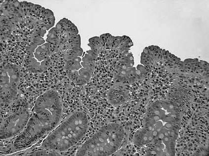
A 6-year-old child presents with diarrhea, malabsorption, and steatorrhea. A photomicrograph from a small intestinal mucosal biopsy is displayed in below figure. An appropriate treatment would be which of the following?
A. alpha-interferon therapy
B. antineoplastic drugs
C. initiation of a gluten-free diet
D. referral to hospice for supportive care
E. surgical resection of a segment of small Bowel
Correct Answer: C
Section: Pathology and Path physiology The child has celiac disease, a disorder resulting from a hypersensitivity reaction to gluten in the diet. Withdrawal of gluten from the diet is usually curative. Clinically, there is diarrhea, malabsorption, and steatorrhea. Histologically, there is villous atrophy of the small intestinal mucosa. A referral to hospice for supportive care (choice D) is unlikely to be necessary; more than 95% of patients respond to the removal of gluten from their diet. Recalcitrant cases are rarely life threatening and may be successfully treated with various forms of hyperalimentation that bypass the small intestine. Because the disease is usually cured by dietary measures, the use of alphainterferon therapy (choice A), antineoplastic drugs (choice B), and surgical resection of the small intestine (choice E) are not appropriate treatment options.
Question 308:
A 56-year-old man complains of increasing dyspnea on exertion over the past few days. He is noted to be overweight and cyanotic. He has smoked cigarettes for at least 35 years and has a long-standing history of persistent cough, producing a large amount of thick mucopurulent sputum. Auscultation reveals scattered rhonchi and wheezes. Histological examination of his lung tissue would most likely how which of the following?
A. expanded alveolar septae infiltrated by mononuclear cells
B. mucous gland hypertrophy and fibrosis of bronchiolar walls
C. neutrophilic exudate occupying the alveoli of an entire lobe
D. pink, proteinaceous layer lining the alveolar spaces
E. thickened basement membranes and many eosinophils
Correct Answer: B
Section: Pathology and Path physiology This presentation is fairly typical for a patient with chronic bronchitis. Microscopically one would expect to see hypertrophy of bronchial mucous glands accompanied by chronic inflammation and fibrosis of bronchiolar walls. Expanded or thickened alveolar septae infiltrated by mononuclear cells (choice A) would be seen when there is an interstitial response in the lung as, for example, in viral or mycoplasmal pneumonia. Neutrophilic exudate occupying the alveoli of an entire lobe (choice C) is a description of a classic lobar pneumonia. Pink, proteinaceous layer lining the alveolar spaces (choice D) is a hyaline membrane that one might see in shock lung or respiratory distress syndrome. Thickened basement membranes and many eosinophils (choice E) would be seen in asthma.
Question 309:
A 10-year-old boy is brought to a pediatric clinic by his mother, who states that he has been voiding brown-colored urine for the past 2 days. The child's history is negative except for a sore throat about 2 weeks ago. Physical examination reveals moderate periorbital edema and mild hypertension. Urinalysis demonstrates red cells, both red and white cell casts, and 2+ protein. If a renal biopsy were obtained, what would be the characteristic light and/or electron microscopic finding in the glomeruli?
A. hypercellularity with duplication of the basement membrane
B. hypercellularity with subepithelial "humps"
C. hypercellularity with "wire loop"lesions
D. normocellularity with effacement of epithelial cell foot processes
E. normocellularity with segmental sclerosis
F. normocellularity with thickened basement membranes
Correct Answer: B
Section: Pathology and Path physiology Acute postinfectious (poststreptococcal) glomerulonephritis occurs more commonly in young children following a sore throat or, less frequently, a skin infection. They usually present with mild periorbital edema, mild to moderate hypertension, and urinary changes. Urinalysis will show hematuria, mild proteinuria, and red and white cells casts. Light microscopy of a renal biopsy will reveal hypercellularity due to proliferation of endothelial and mesangial cells and the infiltration of neutrophils and monocytes. Immune complexes are deposited in the subepithelium of the glomerular basement membrane and may be seen as subepithelial "humps" by electron microscopy. Choices A, C, D, E, and F refer to the microscopic findings in membranoproliferative glomerulonephritis type 1, lupus nephritis, nil disease, focal segmental glomerulosclerosis, and membranous glomerulopathy, respectively.
Question 310:
Exhibit: Please refer to the exhibit. A 14-month-old baby boy is brought to your office by his mother because he seems to be in pain whenever he tries to move. During your physical examination you note bowing of his legs, depression of the sternum with outward projection of the ends of the ribs, reluctance to move his limbs, and numerous bruises on his legs as well as gingival hemorrhages. These findings lead you to suspect that this child suffers from a dietary deficiency of which of the following vitamins?
A. A
B. B
C. C
D. D
E. E
F. F
Correct Answer: D
Section: Pathology and Path physiology Deficiencies of vitamin C and vitamin D can produce similar skeletal abnormalities in young children such as those listed. However, a major difference is that vitamin C deficiency is accompanied by hemorrhages, as seen in this child. This also leads to hemarthrosis (bleeding into joints) that makes movement very painful. Vitamin A deficiency (choice A) is associated with night blindness, with or without keratomalacia and papular dermatitis. Vitamin B1 deficiency (choice B) produces beriberi marked by polyneuropathy, heart failure, and edema (or Wernicke syndrome in chronic alcoholics). Vitamin B12 deficiency (choice C) produces megaloblastic anemia and subacute combined degeneration of the spinal cord. Vitamin D deficiency (choice E) produces osteomalacia in adults and rickets in children due to defective mineralization of bone. Vitamin K deficiency (choice F) can result in a bleeding diathesis because it is required for the activity of clotting factors II, VII, IX, and X.
Nowadays, the certification exams become more and more important and required by more and more enterprises when applying for a job. But how to prepare for the exam effectively? How to prepare for the exam in a short time with less efforts? How to get a ideal result and how to find the most reliable resources? Here on Vcedump.com, you will find all the answers. Vcedump.com provide not only USMLE exam questions, answers and explanations but also complete assistance on your exam preparation and certification application. If you are confused on your USMLE-STEP-1 exam preparations and USMLE certification application, do not hesitate to visit our Vcedump.com to find your solutions here.