A neurologist is performing the neurological examination on a patient who recently suffered a head trauma. You note that, as part of the examination, she uses a cotton swab to touch the upper part of the auricle, the external auditory meatus, and the lobule. The external auditory meatus of the ear is innervated by which of the following?
A. vagus (tenth cranial) nerve
B. great auricular nerve
C. auriculotemporal nerve
D. greater occipital nerve
E. facial (seventh cranial) nerve
Correct Answer: A
Section: Anatomy The vagus (tenth cranial) nerve innervates the external auditory meatus of the ear. The great auricular nerve (choice B) innervates the lobule of the auricle and the auriculotemporal nerve (choice C), the superior aspect of the auricle. In fact, a sensory test which includes these three parts of the ear tests the integrity of the trigeminal (fifth cranial) nerve by the auriculotemporal nerve, the vagus (tenth cranial) nerve by its branch innervating the auditory meatus, and spinal nerves C2-3 by their great auricular branch. The test thus covers the upper and lower medulla and the upper spinal cord. The greater occipital nerve (choice D) is a branch of the cervical plexus originating from C2 and innervates the scalp of the back of the head. The facial (seventh cranial) nerve provides only motor innervation to the face and scalp areas.
Question 832:
In a medial medullary syndrome that involves a left-sided branch of the anterior spinal artery, which of the following deficits is seen?
A. deviation of the tongue to the left, hemiplegia of arm and leg on the left
B. deviation of the tongue to the right, hemiplegia of arm and leg on the right
C. loss of conscious proprioception and precise tactile discrimination over the right side of the body exclusive of the face
D. only deviation of the tongue to the left
E. only hemiplegia on the right
Correct Answer: C
Section: Anatomy A vascular lesion affecting the left caudal medulla involves the left medial lemniscus, left hypoglossal nerve fibers, and the left medullary pyramid. Involvement of the left medial lemniscus produces somatosensory deficits involving the right side of the body. Damage to the left hypoglossal nerve would result in deviation of the protruded tongue to the left (and other lower motoneuron signs), and damage to the left pyramid results in right hemiplegia (choices A and B involve incorrect combinations) along with other upper motoneuron signs. Choices D and E are incorrect because they fail to combine involvement of the tongue and contralateral hemiplegia.
Question 833:
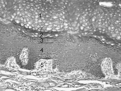
During a routine physical examination, you notice that your patient, a 35-year-old avid surfer, has spots of abnormal pigmentation on two of her fingers. You explain to your patient that long-term exposure to the sun increases the risk of neoplastic changes and that you would like to perform biopsies to verify the nature of the abnormal pigmentation. Referring to following figure, cells from which layer of the epidermis are most vulnerable to neoplastic changes due to long-term exposure to the sun?
A. 1
B. 2
C. 3
D. 4
E. 5
Correct Answer: D
Section: Anatomy Long-term exposure to the sun increases the risk of alteration of the DNA structure by cleavage, ionizing radiation, or recombination of DNA with highly reactive free radicals. These changes can result in neoplastic changes or death in skin cells. In the skin, mitosis occurs only in the malpighian layer formed by the stratum basale and the stratum spinosum of the epidermis. The DNA of dividing cells is more vulnerable to the harmful effects of the sun, and neoplastic changes are usually observed in the Malpighian layer. They are not seen in the stratum corneum (choice A), stratum lucidum (choice B), or stratum granulosum (choice C). Choice E represents the dermis located below the epidermis, which is the only skin layer considered in this question.
Question 834:
A female 44-year-old patient suffers from acute bacterial sinusitis localized to the frontal sinus. The patient
displays a mucopurulent greenish discharge from the nose bilaterally, with associated fever and malaise.
The patient also complains of pain over the forehead with headache.
Which of the following innervates the frontal sinus?
A. anterior ethmoidal nerve
B. lacrimal nerve
C. nasociliary nerve
D. posterior ethmoidal nerve
E. supraorbital nerve
Correct Answer: E
Section: Anatomy The frontal sinus is innervated by the supraorbital and supratrochlear branches of the frontal nerve. All nerves mentioned in this question are branches of ophthalmic division (V1) of the trigeminal (fifth cranial) nerve. The anterior (choice A) and the posterior (choice D) ethmoidal nerves arise from the nasociliary nerve (choice C). They innervate the ethmoid and sphenopalatine sinuses. The lacrimal nerve (choice B) carries in its terminal segment the parasympathetic innervations to the lacrimal gland and provides sensory innervation to the upper eyelid.
Question 835:
Occlusion of which of the following vessels affects the entire dorsolateral part of the rostral medulla (level of the restiform body) and produces the lateral medullary (Wallenberg) syndrome?
A. anterior inferior cerebellar artery
B. anterior spinal artery
C. posterior inferior cerebellar artery
D. posterior spinal artery
E. superior cerebellar artery
Correct Answer: C
Section: Anatomy The posterior inferior cerebellar artery supplies the rostral, dorsolateral medulla. The posterior spinal (choice D) and anterior spinal (choice B) arteries supply dorsal and ventral portions, respectively, of the caudal medulla. The anterior inferior cerebellar (choice A) and superior cerebellar (choice E) arteries supply portions of the pons and mesencephalon.
Question 836:
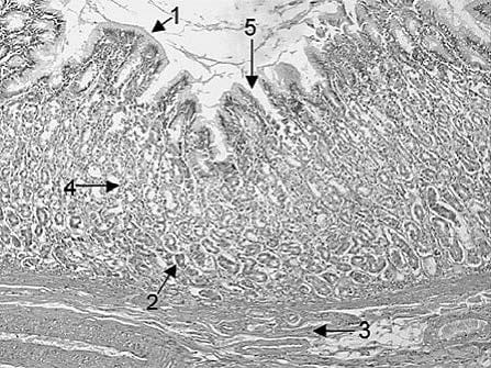
The chief or peptic (zymogenic) cells of the gastric glands secrete pepsinogen. The latter is converted to pepsin, a 35-kilodalton (kDa) proteolytic enzyme, when the pH in the stomach falls below 5.0. In Following figure, which of the following arrows point to the location of chief or peptic (zymogenic) cells?
A. 1
B. 2
C. 3
D. 4
E. 5
Correct Answer: B
Section: Anatomy Arrow 2 points to the base of the gastric glands where chief or peptic (zymogenic) cells tend to be clustered. Arrow 1 points to the luminal surface of the stomach where mucus-secreting cells are found. Arrow 3 points to the muscularis mucosae. Arrow 4 points to the middle of the gastric glands where parietal or oxyntic cells tend to be most numerous. Arrow 5 point to the side of a gastric pit where mucus-secreting cells are also found.
Question 837:
A 62-year-old patient diagnosed with prostate carcinoma complains of a right-sided headache worsening over 4 days and displays a drooping right upper eyelid. Examination reveals a right third nerve palsy. An MRI reveals a single metastasis of the prostatic carcinoma in the right side of the midbrain, causing Benedikt's syndrome. Which of the following signs would also be seen in this patient?
A. complete paralysis of facial expression musculature on the left side
B. deviation of the tongue to the right
C. intention tremor in the left upper and lower extremity
D. ipsilateral hemiplegia
E. vertical gaze palsy
Correct Answer: C
Section: Anatomy Benedikt's syndrome results from a lesion situated in the tegmentum of the midbrain, at the level of the third cranial nerve (oculomotor) nucleus and its associated tracts, as exemplified by ptosis and third nerve palsy in this patient. The red nucleus is also affected at this level giving rise to motor impairment displayed by the intention tremor. Since the rubrospinal tract crosses at the level of the midbrain to project to the opposite side of the body, the tremor will manifest itself contralateral to the side to the lesion. The seventh cranial nerve (facial) nucleus is located in the pons, and the facial musculature (choice A) in this patient would not be affected. Likewise, the twelfth cranial nerve (hypoglossal) nucleus is located in the medulla, and the innervation of the tongue (choice B) would be spared in this patient. A lesion causing a pure Benedikt's syndrome would be confined to the midbrain tegmentum and not affect the corticospinal tract. Ipsilateral hemiplegia (choice D) would not be present in this patient. Finally, vertical gaze palsy (choice E) results from a lesion or compression of the midbrain tectum and not of the tegmentum.
Question 838:
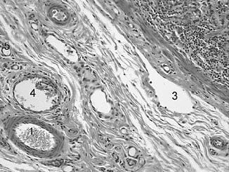
Clinical edema results when lymphatic vessels are blocked or when the volume of extracellular fluid exceeds the drainage capacity of the lymphatic vessels. Which of the following numbered structures in following figure is a lymphatic vessel?
A. 1
B. 2
C. 3
D. 4
E. 5
Correct Answer: C
Section: Anatomy An irregular outline, a thin wall, and the lack of erythrocytes in the lumen characterize lymphatic vessel. Arterioles (choices A and B) have thicker walls and contain erythrocytes. Venules (choice D) are thin-walled but they contain erythrocytes. Capillaries (choice E) are small in diameter and they contain erythrocytes.
Question 839:
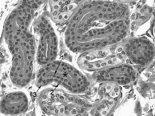
The histological structure marked by the asterisk in Fig. 1-4 is which of the following structures from the integumentary system?
A. aprocrine sweat gland
B. dermal papilla
C. eccrine sweat gland
D. hair follicle
E. sebaceous gland
Correct Answer: C
Section: Anatomy This is the secretory portion of the eccrine sweat gland, recognizable by its three cell types. The apical dark cells (arrow 1) are closest to the lumen. The clear or basal cells (arrow 2) and the myoepithelial cells (arrow 3) are located against the basal lamina. Characteristically, these cells are large and the lumen is small. The apocrine sweat gland (choice A) is lined with simple cuboidal epithelium and thus has a large lumen. The dermal papilla (choice B) is formed by fibroblasts, not epithelia. The hair follicle (choice D) is formed by three concentric zones of keratinized cells and does not have a lumen. The sebaceous glands (choice E) are appendages of the hair follicle and their lumen is lined by stratified squamous epithelium.
Question 840:
About 75% of the blood supply of the spinal cord is derived from the anterior spinal artery. This artery arises from which of the following?
A. artery of Adamkiewicz
B. basilar artery
C. internal carotid artery
D. posterior inferior cerebellar artery
E. vertebral artery
Correct Answer: E
Section: Anatomy At the level of the foramen magnum, the bilateral vertebral arteries supply a medial branch each and they combine to form the anterior spinal artery. As the anterior spinal artery continues caudally, it is supplied by radicular arteries arising from the aorta and its branches. The artery of Adamkiewicz (choice A) is the largest radicular artery (arteria radicularis magna), frequently arising from a segmental branch of the thoracic aorta at the level of T10. The basilar artery (choice B) is formed by the joining of the vertebral arteries superior to the foramen magnum and thus does not provide blood supply to the spinal cord. Its branches supply the brainstem. The internal carotid artery (choice C) supplies the orbit via its ophthalmic branch and the brain by its anterior and middle cerebral branches. The posterior inferior cerebellar artery (choice D) is a branch of the vertebral artery, providing vascular supply to the medulla oblongata and the inferior aspect of the cerebellum.
Nowadays, the certification exams become more and more important and required by more and more enterprises when applying for a job. But how to prepare for the exam effectively? How to prepare for the exam in a short time with less efforts? How to get a ideal result and how to find the most reliable resources? Here on Vcedump.com, you will find all the answers. Vcedump.com provide not only USMLE exam questions, answers and explanations but also complete assistance on your exam preparation and certification application. If you are confused on your USMLE-STEP-1 exam preparations and USMLE certification application, do not hesitate to visit our Vcedump.com to find your solutions here.