The protein encoded by the APC gene is a tumor suppressor the role of which is to regulate the activity of cellular signaling induced by the Wnt growth factor. Therefore, loss of APC activity is associated with unrestrained cellular proliferation. Which of the following signaling molecules is the target for APC interaction?
A. beta-catenin
B. MYC
C. p53
D. pRB
E. von Hippel-Lindau protein (pVHL)
Correct Answer: A
Section: Biochemistry The protein of the Wnt signaling cascade, to which APC associates, is beta-catenin. Using antibodies specific for the NH2-terminus of APC, it was possible to coprecipitate additional PCassociated proteins leading to the identification that one of these APC-associated proteins was beta-catenin. The catenins are a family of proteins that interact with the cytoplasmic portion of the cadherins (cell-cell adhesion family of proteins), thus linking the cadherins to the actin cytoskeleton. Catenins are equally important in the signaling cascade initiated by the Wnt family of proteins that are involved in embryonic patterning, development of the nervous system. The Wnt proteins are secreted factors that interact with cell-surface receptors. Wntreceptor interaction induces the activity of the cytoplasmic phosphoprotein dishevelled. Activated dishevelled inhibits the serine/ threonine kinase glycogen synthase kinase-3- beta (GSK-3). When GSK-3-beta is inhibited, beta-catenin becomes hypophosphorylated. The hypophosphorylated form of beta-catenin migrates to the nucleus and interacts with transcription factors, thereby, inducing expression of various genes. The role of APC in this pathway is to bind phosphorylated beta-catenin. The APC-beta-catenin complex stimulates the breakdown of beta-catenin. Therefore, mutations that lead to a loss of APC, or to a loss of the portion of the APC protein that interacts with beta-catenin, would lead to constitutive activation of several genes that could then promote the transformed phenotype. None of the other proteins (choices BE) are known to interact with APC.
Question 472:
Metformin is one of the most prescribed hypoglycemia-inducing drugs in the treatment of Type II diabetes. One of the effects of metformin is a reduction in adipose tissue lipolysis, which is effected via the activation of AMP-activated kinase (AMPK). Which of the following actions of AMPK explains the adipose tissue benefits of metformin?
A. activation of ACC
B. activation of FAS
C. inhibition of hormone-sensitive lipase
D. inhibition of mammalian target of rapamycin (mTOR)
E. inhibition of 6-PFK-2
Correct Answer: C
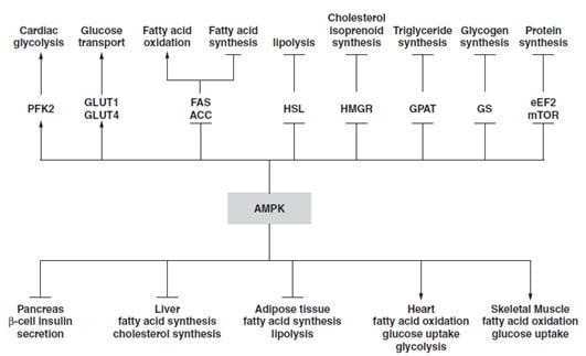
Section: Biochemistry AMP-activated protein kinase (AMPK) was first discovered as an activity that inhibited preparations of ACC and 3-hydroxy-3-methylglutaryl- CoA reductase (HMG-CoA reductase, HMGR) and was induced by AMP. AMPK induces a cascade of events within cells in response to the ever changing energy charge of the cell. The role of AMPK in regulating cellular energy charge places this enzyme at a central control point in maintaining energy homeostasis (see below figure). Once activated, AMPK- ediated phosphorylation events switch cells from active ATP consumption (e.g., fatty acid and cholesterol biosynthesis) to active ATP production (e.g., fatty acid and glucose oxidation). Other important activities attributable to AMPK are regulation of insulin synthesis and secretion in pancreatic islet beta-cells. As shown in AMPK inhibits (not activates) both ACC (choice A) and FAS (choice B). Activation (not inhibition) of PFK-2 (choice E) occurs in response to AMPK. Although AMPK does indeed inhibit mTOR (choice D), this inhibition does not have any influence on adipose tissue lipolysis.
Question 473:
Regulation of cholesterol, fatty acid, phospholipid, and triglyceride synthesis is controlled in part by the level of sterols. These sterol-regulated biosynthesis pathways involve a unique family of transcription factors that are best described by which of the following?
A. Binding of sterols to these factors induces a conformational change preventing interaction with RNA polymerase.
B. They are membrane-bound proteins and only released on activation of a sterolregulated protease.
C. They contain zinc fingers that bind sterols leading to their activation.
D. They require interaction with sterols, much like steroid hormone receptors, prior to binding to DNA.
E. On sterol binding, these factors are recognized by the proteosome and thus targeted for degradation.
Correct Answer: B
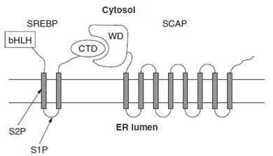
Section: Biochemistry The continual alteration of the intracellular sterol content occurs through the regulation of key sterol synthetic enzymes, as well as by altering the levels of cell-surface LDL receptors. As cells need more sterol they will induce their synthesis and uptake, conversely when the need declines synthesis and uptake are decreased. Regulation of these events is brought about primarily by sterol-regulated transcription of key rate-limiting enzymes and by the regulated degradation of HMGR. Activation of transcriptional control occurs through the regulated cleavage of the membrane- bound transcription factor SREBP (there are three SREBPs). Sterol control of transcription affects more than 30 genes involved in the biosynthesis of cholesterol, triacylglycerols, phospholipids, and fatty acids. Transcriptional control requires the presence of an octamer sequence in the gene termed the sterol regulatory element, SRE-1. It has been shown that SREBP is the transcription factor that binds to SRE- 1 elements. All three SREBPs are proteolytically regulated by sterols. Full-length SREBPs have several domains and are embedded in the membrane of the ER. The N-terminal domain contains a transcription factor motif of the basic helix-loop-helix (bHLH) type that is exposed to the cytoplasmic side of the ER (see below figure). When sterols are scarce, cleavage of the full-length SREBP takes place with the result being that the N-terminal bHLH motif is released into the cytosol. The bHLH domain then migrates to the nucleus to direct transcription. Conversely, when sterols are abundant, cleavage of SREBP is inhibited. To control the level of SREBP-mediated transcription, the soluble bHLH domain is itself subject to rapid proteolysis. The cleavage of SREBP is carried out by two distinct enzymes, one of which is regulated by sterols. The regulated cleavage occurs in the lumenal loop between the two transmembrane domains. This cleavage is catalyzed by site-1 protease, S1P. High sterol content blocks the activity of S1P. The second cleavage, catalyzed by site-2 protease, S2P, occurs in the first transmembrane span, leading to release of active SREBP. In order for S2P to act on SREBP, site-1 must already have been cleaved. Additional studies on sterol-regulated gene expression demonstrated that cleavage of SREBP by S1P is controlled by the level and action of an additional protein termed, SREBP leavage-activating protein, SCAP.
SCAP is a large protein also found in the ER membrane and contains at least eight transmembrane spans. The C-terminal portion, which extends into the cytosol, has been shown to interact with the C- terminal domain of SREBP. This C-terminal region of SCAP contains four motifs called WD40 repeats. The WD40 repeats are required for interaction of SCAP with SREBP. The function of SCAP is to positively stimulate S1P-mediated cleavage of SREBP. The function of sterols is to inhibit this positive action of SCAP. The activity of SCAP involves movement from the ER to the Golgi and back. Because the C-terminus of SCAP interacts with SREBP, movement of SCAP takes SREBP along for the ride. When sterols are low, SCAP and SREBP move to the Golgi. This transit is required for SREBP cleavage as S1P is Golgi-localized. When sterols are high, movement of SCAP is halted. Thus, the overall effect of sterols is to regulate the ability of SCAP to present SREBP to S1P. None of the other choices (A, CE) represent the mechanisms of sterol-mediated regulation of gene expression.
Question 474:
Regulation of iron homeostasis occurs by controlling the amount that circulates in the serum, as well as the amount contained within cells. One mechanism that plays a role in this homeostasis is iron- mediated control of the level of the intracellular iron-binding protein ferritin. Which of the following represents the mechanism of iron regulation of ferritin levels?
A. Binding of iron to ferritin leads to secretion of the complex from cells and subsequent excretion in the urine.
B. Ferritin exists as a tetramer and when iron binds, the affitinity for additional iron atoms increases.
C. Iron binds an additional protein that acts as a regulator of ferritin mRNA translation, high iron leads to increased translation and thus increased ironbinding capacity.
D. When excess iron binds to ferritin, it decreases the half-life of the protein allowing the iron to be released to the plasma and excreted.
Correct Answer: C
Question 475:
Which of the following factors of blood coagulation is the major inhibitor of the extrinsic clotting cascade?
A. antithrombin III
B. high molecular weight kininogen
C. lipoprotein-associated coagulation factor
D. alpha-2-macroglobulin
E. protein C
Correct Answer: C
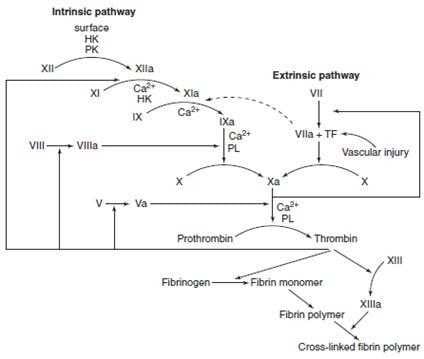
Section: Biochemistry The major mechanism for the inhibition of the extrinsic pathway occurs at the tissue factor--factor VIIa-Ca2 +-Xa--complex. The protein, lipoprotein-associated coagulation inhibitor (LACI, formerly named anticonvertin), specifically binds to this complex. LACI is composed of three tandem protease inhibitor domains. Domain 1 binds to factor Xa and domain 2 binds to factor VIIa only in the presence of factor Xa. Antithrombin III (choice A) is the most important of four thrombin regulatory proteins. This is because antithrombin III can also inhibit the activities of factors IXa, Xa, XIa, and XIIa. The activity of antithrombin III is potentiated in the presence of heparin. Heparin binds to a specific site on antithrombin III, producing an altered conformation of the protein, and the new conformation has a higher affinity for thrombin as well as its other substrates. This effect of heparin is the basis for its clinical use as an anticoagulant. The naturally occurring heparin activator of antithrombin III is present as heparan and heparan sulfate on the surface of vessel endothelial cells. It is this feature that controls the activation of the intrinsic coagulation cascade. HMWK (choice B) is important for initiation of the intrinsic pathway. When prekallikrein, HMWK, factor XI, and factor XII are exposed to a negatively charged surface, they become active. This is termed the contact phase. Exposure of collagen to a vessel surface is the primary stimulus for the contact phase. In addition to antithrombin III, thrombin activity is also inhibited by alpha-2-macroglobulin (choice D). Protein C (choice E) is activated by thrombin when thrombin is bound to thrombomodulin. Active protein C functions with its cofactor, protein S, to degrade factors VIIIa and Xa.
Question 476:
Mutant genes that affect several organ systems and bodily functions frequently show variable expressivity, a phenomenon referred to as phenotypic variation. The most striking example of phenotypic variability is manifest in an autosomal- dominant condition characterized by the appearance of café au lait spots on the skin and cutaneous and subcutaneous neurofibromas. These symptoms are associated with which of the following?
A. familial adenomatous polyposis
B. FH
C. Li Fraumeni syndrome
D. von Hippel-Lindau syndrome
E. von Recklinghausen disease (type I neurofibromatosis)
Correct Answer: E
Question 477:
Exhibit:
Please refer to the exhibit. When fatty acids with odd numbers of carbon atoms are oxidized in the beta-oxidation pathway the final product is 1 mole of acetyl- CoA and 1 mole of the 3-carbon molecule, propionyl-CoA. In order to use the propionyl carbons, the molecule is carboxylated and converted ultimately to succinyl-CoA and fed into the TCA cycle. Which of the following represents the vitamin cofactor required in one of the steps of this conversion?
A. A
B. B
C. C
D. D
E. E
Correct Answer: A
Section: Biochemistry Propionyl-CoA is converted to succinyl- CoAin a series of reactions using three different enzymes. It is first carboxylated in an ATPdependent reaction catalyzed by propionyl- CoA carboxylase, an enzyme that requires biotin as a cofactor. The product of the first reaction, d-methylmalonyl-CoA is then converted to lmethylmalonyl-CoA by methylmalonyl-CoA racemase. Finally, methylmalonyl-CoA is converted to succinyl-CoA by the cobalaminrequiring enzyme, methylmalonyl-CoA mutase. None of the other vitamins (choice B, C, D, and E) are required in this pathway.
Question 478:
Many effective anticancer drugs function as such by interfering with processes of DNA replication. The drug, doxorubicin, is useful in the treatment of lymphomas and breast cancers because of its ability to interfere with which of the following enzyme activities?
A. DNA ligase
B. DNA polymerase-alpha
C. primase
D. topoisomerase II
E. uracil N-glycosylase
Correct Answer: D
Section: Biochemistry Doxorubicin is a drug of the class that functions by interfering with topoisomerases. In particular, doxorubicin inhibits topoisomerase II. During the process of DNA replication ,the two strands of the DNA helix are separated. As the replication fork progresses toward sites of chromosomal attachment to the scaffold, there is an increase in the torsional stress on the helix due to supercoiling. These torsional stresses are relieved by the action of topoisomerases that introduce nicks into the DNA, which allows the strands to unwind the supercoils. Topoisomerase I introduces single-strand nicks, whereas topoisomerase II introduces double-strand nicks. Interference with the action of topoisomerases would thus impair the rate of DNA replication and this would be detrimental for rapidly proliferating cells such as cancer. Doxorubicin does not affect the activity of any of the other enzymes (choices AC, E).
Question 479:
Which of the following clotting factors forms an active complex with tissue factor (factor III) to initiate the extrinsic clotting cascade?
A. factor I (fibrinogen)
B. factor VII (proconvertin)
C. factor VIII
D. factor IX
E. protein S
Correct Answer: B
Section: Biochemistry
The extrinsic pathway (see below figure)
is initiated at the site of injury in response to the release of tissue factor (factor III). Tissue factor is a cofactor in the factor VIIa-catalyzed (lower case alpha refers to the active form of the coagulation factors) activation of factor X. The formation of a complex between factor VIIa and tissue factor is believed to be a principal step in the overall clotting cascade. Evidence for this stems from the fact that persons with hereditary deficiencies in the components of the contact phase of the intrinsic pathway do not exhibit clotting problems. Factor VIIa, a gamma-glutamate (gla residue) containing serine protease, cleaves factor X to factor Xa in a manner identical to that of factor IXa of the intrinsic pathway. The activation of factor VII occurs through the action of thrombin or factor Xa. The ability of factor Xa to activate factor VII creates a link between the intrinsic and extrinsic pathways. An additional link between the two pathways exists through the ability of tissue factor and factor VIIa to activate factor IX. Fibrinogen (choice A) is cleaved by thrombin (factor IIa), yielding fibrin monomers that polymerize to form a fibrin clot. The activation of factor VIII (choice C) to factor VIIIa occurs in the presence of minute quantities of thrombin. Factor VIIIa is required for the activation of factor X, which represents the convergence of the intrinsic and extrinsic pathways. The activation of factor X to Xa requires assemblage of the tenase complex (C and factors VIIIa, IXa, and X) on the surface of
activated platelets. As the concentration of thrombin increases, factor VIIIa is ultimately cleaved by thrombin and inactivated. This dual action of thrombin on factor VIII acts to limit the extent of tenase complex formation, and thus the extent of the coagulation cascade. Factor IX choice D) functions only in the intrinsic cascade. When activated, factor IXa aids in the activation of factor X by functioning in the tenase complex as described above. Protein S (choice E) is a cofactor for protein C. Protein C is itself activated by thrombin and when active, cleaves factors VIIIa and Va, thus limiting the extent of the coagulation cascade.
Question 480:
Which of the following familial cancers results from a defect in the tumor suppressor gene, p53?
A. familial adenomatous polyposis coli (APC)
B. Li Fraumeni syndrome
C. neurofibromatosis type 1
D. retinoblastoma
E. Wilms tumor
Correct Answer: B
Section: Biochemistry Numerous cancers are caused by the loss of function of tumor suppressor genes. LFS is a rare form of inherited cancer that involves breast and colon carcinomas, soft-tissue sarcomas, osteosarcomas, brain tumors, leukemia, and adrenocortical carcinomas. These tumors develop at an early age in LFS patients. The tumor suppressor gene found responsible for LFS is p53. Mutant forms of p53 are found in approximately 50% of all tumors. The normal p53 protein functions as a transcription factor that can induce either cell-cycle arrest or apoptosis (programmed cell death) in response to DNA damage. FAP (choice A) is a rare inherited form of colon cancer. Germline mutations in the APC gene are responsible for FAP. All cases of neurofibromatosis (choice C) arise by inheritance of a mutant allele. Roughly 50% of all affected individuals carry new mutations, which appear to arise paternally, possibly reflecting genomic imprinting. The gene responsible for type 1 neurofibromatosis is termed NF1. Germline mutations at the NF1 locus result in multiple abnormal melanocytes 168 3: Biochemistry (caféau lait spots) and benign neurofibromas. Some patients also develop benign pheochromocytomas and CNS tumors. Asmall percentage of patients develop neurofibrosarcomas, which are likely to be Schwann cell derived. Retinoblastoma (choice D) is a tumor of retinal cells, which develops in children between birth and 4 years of age. The gene responsible is termed the retinoblastoma susceptibility gene (RB) and the protein product pRB. Wilms' tumor (choice E) is a form of childhood kidney cancer. The gene responsible for this disease has been identified and is called WT1 (Wilms' tumor 1).
Nowadays, the certification exams become more and more important and required by more and more enterprises when applying for a job. But how to prepare for the exam effectively? How to prepare for the exam in a short time with less efforts? How to get a ideal result and how to find the most reliable resources? Here on Vcedump.com, you will find all the answers. Vcedump.com provide not only USMLE exam questions, answers and explanations but also complete assistance on your exam preparation and certification application. If you are confused on your USMLE-STEP-1 exam preparations and USMLE certification application, do not hesitate to visit our Vcedump.com to find your solutions here.