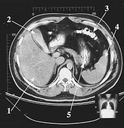
A 42-year-old female patient has to undergo emergency cholecystectomy due to intense biliary colic. The structure to be removed during the surgery is indicated in following figure by which of the following arrows?
A. 1
B. 2
C. 3
D. 4
E. 5
Correct Answer: B
Section: Anatomy Arrow 2 points to the gallbladder, which will be removed during the cholecystectomy (surgical removal of the gallbladder). Biliary colic may be due to impaction of a gallstone in the cystic duct, resulting in cholecystitis (inflammation of the gallbladder). Arrow 1 (choice A) points to the liver. Arrow 3 (choice C) points to the transverse colon. Arrow 4 (choice D) points to the spleen and arrow 5 (choice E) indicates the stomach, recognizable by its internal rugae.
Question 712:
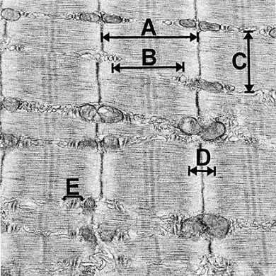
In Fig. following figure which labeled bracket spans a sarcomere?
A. A
B. B
C. C
D. D
E. E
Correct Answer: A
Section: Anatomy
An individual sarcomere, the unit of contraction in striated muscle, spans the interval between successive
Z lines. Each sarcomere encompasses an A-band (choice B) and half of each of two I bands (choice D).
Each myofibril (choice C) of a striated muscle fiber is composed of a tandem series of sarcomeres.
Coupling of excitation and contraction is a critical function of the triad (choice E), which is composed of a T
tubule interposed between two cisternae of the sarcoplasmic reticulum.
Question 713:
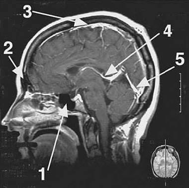
The great cerebral vein of Galen indicated by arrow 4 in following figure is formed by the union of two internal cerebral veins and drains into which of the following?
A. confluence of sinuses
B. frontal sinus
C. sphenoid sinus
D. straight sinus
E. superior sagittal sinus
Correct Answer: D
Section: Anatomy The great cerebral vein of Galen (arrow 4) drains posteriorly into the straight sinus (arrow 5). The union of the superior sagittal sinus (choice E; arrow 3) and the straight sinus forms the confluence of sinuses (choice A). The straight sinus and superior sagittal sinus are dural venous sinuses: they contain venous blood draining form the brain, skull, and scalp. The frontal sinus (choice B) and the sphenoid sinus (choice C) are bony sinuses: they are hollow, airfilled structures and do not drain venous blood.
Question 714:
A 32-year-old female professional gardener complains of increasing muscle weakness and fatigue during the day, requiring her to take frequent rests. She also reports that she cannot enjoy her meals any more because her muscles of mastication quickly weaken and she has to stop chewing. When she watches television at night for a long period of time, her vision becomes blurry and she sees double. Her neurologist makes a preliminary diagnosis of myasthenia gravis. Which of the following is the cause of myasthenia gravis?
A. Acetylcholine synthesis in motor neurons is impaired.
B. Acetylcholinesterase synthesis is inhibited.
C. Autoantibodies destroy cholinergic receptors at the postsynaptic membrane preventing binding of acetylcholine.
D. Neurotransmitter release is impaired at the presynaptic membrane of the neuromuscular junction.
E. Signal transduction within the muscle is impaired.
Correct Answer: C
Section: Anatomy Myasthenia gravis is an autoimmune disorder where autoantibodies target the postsynaptic cholinergic receptors and destroy them. Acetylcholine released from motor neurons is thus unable to bind and the muscle contraction weakens due to decreased neurotransmitter communication. Acetylcholine synthesis in motor neurons (choice A) remains normal. Acetylcholinesterase (choice B) is the enzyme which degrades acetylcholine and its synthesis is not affected in myasthenia gravis. The cholinergic neurotransmitter release mechanism (choice D) at the presynaptic membrane, as well as the signal transduction mechanism (choice E) within the muscle, remain normal.
Question 715:
Lymph nodes are populated by lymphocytes that exit the vascular compartment to gain access to the parenchyma of the node by passing through the walls of which of the following?
A. afferent lymphatic vessels
B. arterioles
C. efferent lymphatic vessels
D. high endothelial postcapillary venules
E. medullary sinuses
Correct Answer: D
Section: Anatomy High endothelial venules (HEV), located primarily in the deep cortex, are specialized to recruit circulating lymphocytes from the blood. Lymphocytes in the circulating blood adhere to the lining endothelial cells of HEV by way of an integrin-based recognition. Lymphocytes then gain access to the lymph node tissue by actively migrating (a process called diapedesis) between or through endothelial cells. Afferent lymphatic vessels (choice A) conduct lymph, not blood, into the lymph node. The source of the lymph is either upstream lymph nodes or tissue fluid from the region supplied by the node. This component of the system serves as a filter and as a mechanism for antigen-presenting cells to enter the node. Arterioles (choice B) are a component of the circulation of the lymph node, but they are not permeable to cell traffic. Efferent lymphatic vessels (choice C) conduct lymph and cells from the lymph node to either the blood circulation or downstream lymph nodes. Lymph in efferent lymphatic vessels conveys immunoglobulins and recirculating lymphocytes to the bloodstream. Medullary sinuses (choice E) are part of a system of passages that filter lymph and direct it from the afferent lymphatic vessels to the efferent lymphatic vessels. Medullary sinuses occupy spaces between medullary cords, which are occupied by large numbers of plasma cells, the cells that secrete immunoglobulins.
Question 716:
In adults, lack of vitamin D gives rise to the disease osteomalacia characterized by progressive softening and bending of the bones. This is due to a defect in the mineralization of the osteoid. Under normal conditions, the osteoid is found along which of the following locations?
A. the interface between osteocytes and bones
B. the interface between osteoprogenitor cells and bone marrow
C. the interface between the fibroblasts in the periosteum
D. the interface between the osteoblast and bone
E. the ruffled border of osteoclasts
Correct Answer: D
Section: Anatomy
Osteoid is the unmineralized organic matrix formed by osteoblasts and found at the interface between
these cells and bone. Osteocytes (choice A) are surrounded by bone and no longer manufacture osteoid.
Osteoprogenitor cells (choice B) are similar to stem cells and do not manufacture bone material.
Fibroblasts (choice C) are cells of connective tissue forming the periosteum and not bone. Osteoclasts
(choice E) are bone-resorbing cells.
Question 717:
Calcitonin decreases blood calcium and boneresorbing activity in which of the following?
A. osteoblast
B. osteoclast
C. osteocyte
D. osteon
E. osteoprogenitor cells
Correct Answer: B
Section: Anatomy Calcitonin reduces the surface ruffling of osteoclasts and their activity. Osteoclasts are formed by fusion of blood monocyte derivatives and are components of the mononuclear phagocyte system. Under the influence of parathyroid hormone, osteoclasts enlarge their ruffled borders and increase their boneresorbing activity. Osteoprogenitor cells (choice E), osteoblast (choice A), and osteocyte (choice C) are bone-forming cells. Osteon (choice D) is another name for the haversian system which is the haversian canal, its contents, surrounding lamellae, and osteocytes.
Question 718:
During development, which of the following structures act as a temporary set of kidneys in the fetus?
A. mesonephroi
B. metanephroi
C. paramesonephric ducts
D. pronephroi
E. ureteric bud
Correct Answer: A
Section: Anatomy The second set or mesonephroi appear late in the fourth week and are functional until the permanent kidneys or metanephroi are fully developed. During development, three sets of kidneys are formed in the embryo. The first set or pronephroi (choice D) are transitional, nonfunctional structures that appear around the fourth week of development. Next come the mesonephroi, then the permanent set of kidneys develops from the metanephroi. The paramesonephric ducts (choice C) are structures developing lateral to the gonads and mesonephric ducts. They play an essential role in the female reproductive system, but are not involved in the formation of the kidneys. The ureteric bud (choice E) is an outgrowth from the mesonephric duct that gives rise to the ureter, renal pelvis, calices, and collecting tubules.
Question 719:
A patient suffering from Charcot-Marie-Tooth disease displays progressive degeneration of peripheral nerves, distal muscle weakness and atrophy, and defects in deep tendon reflexes. This condition is associated with an abnormal mutation in the gene encoding connexin-32. Connexins are normally found in which type of cell junctions?
A. communicating (gap) junction
B. hemidesmosome
C. macula adherens (spot desmosome)
D. occluding (tight) junction
E. zonula adherens (belt desmosome)
Correct Answer: A
Section: Anatomy Communicating (gap) junctions are formed by connexins, which associate together in groups of six to form connexons. The alignment of connexions between two cells allows for direct channels of communication between their cytoplasms, facilitating the transfer of molecules such as calcium or cyclic adenosine monophosphate (cAMP). Hemidesmosome (choice B), macula adherens (spot desmosome; choice C), and zonula adherens (belt desmosome; choice E) are classified as anchoring junctions. They are associated with intermediate filaments (hemidesmosome and macula adherens) or with actin microfilaments (zonula adherens), but not connexins. Occluding (tight) junctions (choice D) contain the proteins occludin and claudin but not connexin.
Question 720:
After removal of cancerous lymph nodes from the lateral pelvic wall, a patient develops painful spasms of the adductor muscles and sensory deficits in the medial thigh region. The adductor muscles are innervated by which of the following nerves?
A. femoral
B. inferior gluteal
C. obturator
D. pudendal
E. sciatic
Correct Answer: C
Section: Anatomy The obturator nerve innervates the adductor muscles and the medial region of the thigh. The nerve originates from the lumbar plexus, runs on the lateral aspect of the pelvic wall, and exits through the obturator canal to reach the medial aspect of the thigh. Lying on the lateral pelvic wall, it may be injured by surgical mishap. The femoral nerve (choice A) innervates the anterior aspect of the thigh and the muscles contained within the sartorius and the quadriceps femoris. The inferior gluteal nerve (choice B) innervates the gluteus maximus muscle and is confined to the gluteal region. The pudendal nerve (choice D) is sensory to the genitalia, motor to the perineal muscles, the external urethral sphincter, and the external anal sphincter. The sciatic nerve (choice E) innervates the hamstring muscles in the posterior aspect of the thigh.
Nowadays, the certification exams become more and more important and required by more and more enterprises when applying for a job. But how to prepare for the exam effectively? How to prepare for the exam in a short time with less efforts? How to get a ideal result and how to find the most reliable resources? Here on Vcedump.com, you will find all the answers. Vcedump.com provide not only USMLE exam questions, answers and explanations but also complete assistance on your exam preparation and certification application. If you are confused on your USMLE-STEP-1 exam preparations and USMLE certification application, do not hesitate to visit our Vcedump.com to find your solutions here.