A 45-year-old female patient develops Parinauds syndrome (vertical gaze palsy) and an MRI shows a large tumor of her pineal gland. The tumor is not only compressing her tectum, causing the vertical gaze palsy, but also obstructing the underlying cerebrospinal fluid pathway causing progressive noncommunicating hydrocephalus. The obstruction causes immediate accumulation of cerebrospinal fluid in which of the following?
A. cerebral aqueduct of Sylvius
B. fourth ventricle
C. lateral ventricle
D. subarachnoid space
E. third ventricle
Correct Answer: E
Section: Anatomy In this situation, the cerebrospinal fluid will accumulate immediately in the third ventricle and cause hydrocephalus. The flow of cerebrospinal fluid is normally from the lateral ventricles (choice C) to the third ventricle and then by way of the cerebral aqueduct of Sylvius (choice A) to the fourth ventricle (choice B). From there the cerebrospinal fluid flows out of the brain into the subarachnoid space (choice D). A blockage of the cerebral aqueduct will result in immediate accumulation of cerebrospinal fluid in the third ventricle. Because the cerebrospinal fluid no longer flows from the ventricular system to the subarachnoid space, the resulting hydrocephalus is termed noncommunicating.
Question 742:
Apatient has been severely injured in the back of the head during a mugging attempt and imaging studies reveal possible fracture of the skull along with the C1 (atlas) vertebra. The patient is also hemorrhaging from the vertebral artery in this location, and the attending surgeon will attempt to stop the bleeding by access through the suboccipital triangle. Which of the following muscles attaches from the transverse process of C1 to the occipital bone and forms the lateral border of the suboccipital triangle?
A. obliquus capitis inferior
B. obliquus capitis superior
C. rectus capitis posterior major
D. rectus capitis posterior minor
E. semispinalis cervicis
Correct Answer: B
Section: Anatomy The obliquus capitis superior, attached between the transverse process of C1 and the suboccipital bone between the nuchal lines. It forms the lateral border of the suboccipital triangle. The obliquus capitis inferior (choice A) is the inferior border of the triangle, attaching from the spinous process of C2 to the transverse process of C1. The rectus capitis posterior major (choice C) inserts from the spinous process of C2 into the inferior nuchal line on the occipital bone; it forms the medial border of the suboccipital triangle. The rectus capitis posterior minor (choice D) lies medial to the rectus capitis posterior major. The semispinalis cervicis (choice E) does not participate in the formation of the suboccipital triangle.
Question 743:
In elderly patients (over 60 years of age), fractures of the neck of the femur following a fall are common. Arterial branches supplying the femoral head and neck are vulnerable to injury during these fractures, and the resulting posttraumatic avascular necrosis affects the head of the femur. In the adult, the most important direct vascular source to the femoral head and neck is which of the following?
A. artery to the head of the femur
B. femoral artery
C. lateral circumflex femoral artery
D. medial circumflex femoral artery
E. superior gluteal artery
Correct Answer: D
Section: Anatomy The medial circumflex femoral artery supplies the most important source of blood to the femoral head and neck. This artery anastomoses with the artery to the head of the femur (choice A), which arises from the obturator artery. However, if the medial circumflex femoral artery is injured, the blood flow in the small artery to the head of the femur may not be sufficient to prevent posttraumatic avascular necrosis of the femoral head. Normally, the medial and lateral circumflex femoral arteries arise from the deep artery of the thigh, but occasionally they arise from the femoral artery (choice B). However, the femoral artery is not a direct vascular source to the head of the femur. The lateral circumflex femoral artery (choice C) and superior gluteal artery (choice E) also supply the hip joint, but their contribution to the head and neck of the femur is less than that of the medial femoral circumflex artery.
Question 744:
A 38-year-old female patient suffers from pleurisy and requires pleural fluid sampling (thoracentesis). The attending physician asks you to perform the procedure at the midaxillary line on the right side. Which of the following would be the appropriate level to perform the procedure?
A. above the level of the 7th rib
B. at the level of the 10th rib
C. at the level of the 5th rib
D. below the level of the 10th rib
E. between the level of the 8th and 10th ribs
Correct Answer: E
Section: Anatomy
The needle for thoracentesis should be inserted in the intercostals spaces between the 8th and 10th ribs.
Remember that the parietal pleura extends approximately two ribs inferior to the lung: at the midaxillary
line, the inferior surface at the lung is at the level of the 8th rib and the parietal pleura at the 10th rib.
Above the level of the 7th (choice A) and 5th (choice C) ribs, the needle will injure the lung. At (choice B)
or below the level of the 10th rib (choice D), it will injure the liver or other abdominal organs.
Question 745:
The sensory innervation of the posterior onethird of the tongue is performed by cranial nerve IX (glossopharyngeal). During development, this region of the tongue develops from which of the following pharyngeal arches?
A. first
B. fourth
C. second
D. sixth
E. third
Correct Answer: E
Section: Anatomy The posterior one-third of the tongue is derived from the third pharyngeal arch and is thus innervated by cranial nerve IX (glossopharyngeal). The first pharyngeal arch (choice A) and second pharyngeal arch (choice C) give rise to the anterior two-thirds of the tongue. The mandibular division of cranial nerve V (trigeminal) provides general sensation to the anterior two-thirds of the tongue, and cranial nerve VII via the chorda tympani provides special sensation (taste). The fourth pharyngeal arch (choice B) gives rise to the epiglottis and, along with the sixth pharyngeal arch (choice D), to the laryngeal cartilages. The nerve to the fourth pharyngeal arch is cranial nerve X (vagus).
Question 746:
The sinoatrial (SA) node initiates the heartbeat by giving off an impulse about 80 times per minute. It is located at the junction of the superior vena cava and right atrium. In about 60% of the cases, the SA node derives its vascular supply from which of the following?
A. anterior interventricular artery
B. left circumflex artery
C. posterior interventricular artery
D. right coronary artery E. right marginal branch
Correct Answer: D
Section: Anatomy In 60% of patients, the right coronary artery supplies the SA node. In a third of the population, the SA node is supplied by the left coronary artery and in some patients it receives branches from both the right and the left. The anterior interventricular (choice A) and left circumflex (choice B) arteries are distal branches of the left coronary artery, too distant to supply the SA node. The right coronary artery normally gives out its SA nodal branch in its proximal portion and then distally gives rise to the right marginal (choice E) and posterior interventricular (choice C) arteries. These are also too distant to supply the SA node.
Question 747:
During surgery to repair a strangulated inguinal hernia, it is discovered that the left testicular vein is thrombosed and must be repaired. The left testicular vein normally drains into which of the following?
A. inferior vena cava
B. left renal vein
C. right common iliac vein
D. right femoral vein
E. right renal vein
Correct Answer: B
Section: Anatomy The left testicular vein drains into the left renal vein. The inferior vena cava (choice A) receives the right testicular vein. The equivalent relationship holds true in the female where the left ovarian vein drains into the left renal vein and the right ovarian vein into the inferior vena cava. The right common iliac vein (choice C), right femoral vein (choice D), and right renal vein (choice E) do not receive gonadal veins.
Question 748:
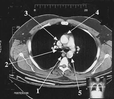
A fourth-year medical student is learning to place a central line. To prepare for this procedure, she reviews X-rays and CT scans in order to gain a proper three-dimensional relationship of the structures involved. In following figure what is the structure pointed to by arrow 1?
A. Ascending aorta
B. Azygos vein
C. Descending aorta
D. Right bronchus
E. Superior vena cava
Correct Answer: E
Section: Anatomy Arrow 1 points to the superior vena cava. The central venous catheter is inserted into the subclavian vein and threaded into the superior vena cava. The ascending aorta (choice A) is labeled by arrow 4 and its counterpart, the descending aorta (choice C) by arrow 5. The azygos vein (choice B) is indicated by arrow
1. Arrow 2 indicates the right bronchus (choice D) just as it leaves the carina.
Question 749:
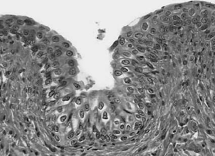
Referring to following figure what is the most likely source of this epithelium?
A. gall bladder
B. salivary duct
C. thick skin
D. trachea
E. urinary bladder
Correct Answer: E
Section: Anatomy The appearance of this epithelium reveals it to be transitional epithelium. It is stratified with a scalloped surface outline. The cells at the bottom layer are cuboidal in appearance and stained darkly; cells in the intermediate layer are polygonal. Cells at the surface of the epithelium are pale-stained, rounded, and large. Transitional epithelium is characteristic of organs of the urinary system, such as the urinary bladder. The gall bladder (choice A) is characterized by simple columnar epithelium. The epithelium of the salivary gland (choice B) is simple cuboidal epithelium. Thick skin (choice C) is made up of stratified squamous epithelium and the trachea (choice D) is lined with pseudostratified columnar epithelium.
Question 750:
Afailure of the truncoconal septum to follow a spiral course results in which of the following conditions?
A. common atrium
B. persistent atrioventricular canal
C. persistent truncus arteriosus
D. Tetralogy of Fallot
E. transposition of the great vessels
Correct Answer: E
Section: Anatomy Transposition of the great vessels occurs when the truncoconal ridges fail to spiral as they divide the outflow tract into two channels. This produces two totally independent circulatory loops with the right ventricle feeding into the aorta and the left ventricle feeding into the pulmonary artery. Common atrium (choice A) results from a complete failure of the septum primum and septum secundum to form. Persistent atrioventricular canal (choice B) results from a failure of the endocardial cushions to fuse and partition the atrioventricular canal into a right and left component. It is accompanied by defects of the atrial and ventricular septa. Persistent truncus arteriosus (choice C) results from a total failure of the truncoconal ridges to develop and partition the outflow tract of the developing heart. Tetralogy of Fallot (choice D) is a related group of defects with the primary malformation being an unequal division of the outflow tract, resulting in pulmonary stenosis. The other eatures of tetralogy are an interventricular septal defect, an overriding aorta, and right ventricular hypertrophy. Survival of the infant depends on the maintenance of a patent ductus arteriosus.
Nowadays, the certification exams become more and more important and required by more and more enterprises when applying for a job. But how to prepare for the exam effectively? How to prepare for the exam in a short time with less efforts? How to get a ideal result and how to find the most reliable resources? Here on Vcedump.com, you will find all the answers. Vcedump.com provide not only USMLE exam questions, answers and explanations but also complete assistance on your exam preparation and certification application. If you are confused on your USMLE-STEP-1 exam preparations and USMLE certification application, do not hesitate to visit our Vcedump.com to find your solutions here.