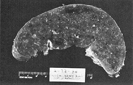
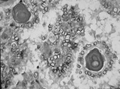
The most likely cause of the pathologic findings in the spleen shown in Figure below is which of the following?
A. amyloidosis
B. metastatic carcinoma
C. septic infarct
D. Hodgkin disease
E. traumatic rupture
Correct Answer: A Section: (none)
Explanation:
Amyloidosis is caused by the deposition of an abnormal proteinaceous material between cells. The majority of the cases are idiopathic, but a small percentage is secondary to chronic infection or inflammation, plasma cell dyscrasias, or immune diseases. One of the characteristic presentations of amyloidosis is splenic infiltration and splenomegaly caused by deposition of amyloid in the follicular regions. Grossly, the spleen has a diffuse, pink, glassy, waxy appearance with obliteration of the white pulp. Amyloid infiltration can also affect the kidneys, liver, and heart. Clinical symptoms are usually due to functional impairment of the diseased organ. The diagnosis of amyloidosis is made by tissue biopsy or, more recently, by fat-pad biopsy looking for amyloid deposits. With Congo red stain, amyloid appears red; with polarization, it shows an apple-green birefringence, which is diagnostic of amyloid.
Question 102:
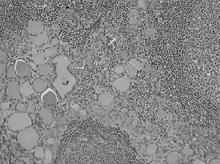
Amiddle-aged female presents with a painless enlargement of the lower aspect of the neck. With appropriate testing this is proven to be thyroid enlargement. Thyroid function tests were normal. Asurgical intervention was performed for diagnostic purposes. Figure below depicts a representative area of how most of thethyroid gland histologic features were seen. What are the clinical thyroid function test characteristics of the last stages of this disease?
A. thyrotoxicosis
B. normal thyroid function tests
C. some degree of hypothyroidism
D. invasion of the recurrent laryngeal nerve
E. hoarseness
Correct Answer: C Section: (none)
Explanation:
Hashimoto thyroiditis is a very common cause of hypothyroidism in the parts of the world where iodine levels are insufficient. The clinical picture is characterized by a gradual enlargement of the thyroid gland with autoimmune destruction. There is a great female to male predominance, with ratio of 10
20:1. Clusters of families are seen which are associated with HLA-DR5 on the major histocompatibility complex (MHC). A few cases are also characterized by HLA-DR3. The pathogenesis is attributed to cellular and humoral immunity which produces thyroid tissue injury. The morphology of the thyroid gland is characterized by typical destruction of the thyroid parenchyma with dense lymphocytic infiltrate and many secondary germinal centers. Occasional scattered Hurthle cells are also seen. Depending on the stage of the disease, extensive areas of fibrosis may also be present. The clinical course of Hashimoto thyroiditis is usually an initial period of time in which the patient may be euthyroid followed by hypothyroidism.
Question 103:
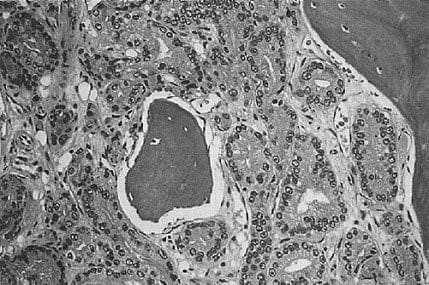
A 60-year-old male is evaluated following a pathologic fracture of his humerus. As part of the workup, a bone biopsy is performed and the photomicrograph is shown in Figure below Which of the following is the most likely diagnosis?
A. benign neoplasm
B. cellular hyperplasia
C. osteogenic sarcoma
D. metastatic lesion
E. chronic leukemia
Correct Answer: D Section: (none)
Explanation:
The photomicrograph accompanying the question shows bone marrow spaces replaced by a well-
differentiated adenocarcinoma. The bone spicules are normal. The glandular structures replacing the
interspicular spaces and replacing the marrow elements are diagnostic of metastatic adenocarcinoma.
Question 104:
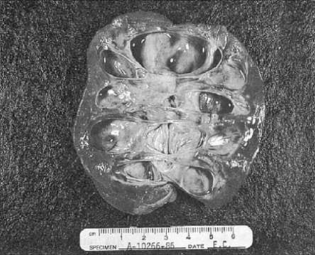
An autopsy was performed following the death of a 72-year-old man. The kidney is shown in Figure below. Which of the following clinical scenarios is most likely to explain the changes seen in this kidney?
A. complete urinary outflow obstruction from an enlarged prostate
B. renal infarct from an embolism originating in the atrium of the heart due to atrial fibrillation
C. long-standing hypertension
D. renal cell carcinoma
E. chronic abuse of analgesics and nonsteroidal anti-inflammatory drugs
Correct Answer: A Section: (none)
Explanation: The photograph that accompanies the question demonstrates severe hydronephrosis, which is due to obstruction of the flow of urine. The obstruction may be located at any site along the urinary outflow tract and may be partial or total, unilateral or bilateral. Because glomerular filtration may continue for some time after the development of the obstruction, the renal pelvis and calices become dilated by continued urine production. The resultant backpressure produces atrophy of the renal parenchyma with obliteration of the pyramids. The degree of hydronephrosis depends on the extent and rapidity of the obstructive process.
Question 105:
A 23-year-old female sought medical help because of a painless asymmetrical enlargement of the lower neck. The patient had no history of dyspnea, dysphagia, hoarseness, or previous radiation exposure. On physical examination, besides the enlarged asymmetrical thyroid gland, there was also a palpable lymphadenopathy. A lymph node biopsysee Figure, was performed. Hematoxilin and eosin (HandE) stained slide shows the lesion.
What are the typical nuclear findings of this tumor?
A. ground glass appearance with intranuclear inclusions
B. abnormal mitosis
C. scant cytoplasm
D. glandular formations
E. squamous metaplasia
Correct Answer: A Section: (none)
Explanation:
Papillary carcinoma of the thyroid is the most common form of thyroid cancer. Most cases are seen between the second and third decade of life and are associated with previous radiation therapy. Many times the first manifestation is a metastasis to the regional neck nodes. The histologic characteristics of papillary carcinoma are branching papillae with single or multiple layers of cuboidal to columnar cells. The characteristic appearance of the nucleus is rather clear, ground-glass (orphan Annie) nuclei. Characteristic intracytoplasmic inclusions, and occasional grooves, are seen. Psammoma bodies are often present in the papillae. The most common variant of papillary carcinoma is the follicular variant, in which the tumor cells form follicular architecture; however, the nuclear changes, as well as focal areas of papillary structures, are enough to make the differential diagnosis from follicular carcinoma.
Question 106:
A 23-year-old female sought medical help because of a painless asymmetrical enlargement of the lower neck. The patient had no history of dyspnea, dysphagia, hoarseness, or previous radiation exposure. On physical examination, besides the enlarged asymmetrical thyroid gland, there was also a palpable lymphadenopathy. A lymph node biopsy (see Figure, was performed. Hematoxilin and eosin (HandE) stained slide shows the lesion.
What is the most appropriate diagnosis?
A. medullary carcinoma of the thyroid
B. follicular carcinoma
C. papillary carcinoma
D. anaplastic carcinoma
E. small cell anaplastic carcinoma
Correct Answer: C Section: (none)
Explanation:
Papillary carcinoma of the thyroid is the most common form of thyroid cancer. Most cases are seen between the second and third decade of life and are associated with previous radiation therapy. Many times the first manifestation is a metastasis to the regional neck nodes. The histologic characteristics of papillary carcinoma are branching papillae with single or multiple layers of cuboidal to columnar cells. The characteristic appearance of the nucleus is rather clear, ground-glass (orphan Annie) nuclei. Characteristic intracytoplasmic inclusions, and occasional grooves, are seen. Psammoma bodies are often present in the papillae. The most common variant of papillary carcinoma is the follicular variant, in which the tumor cells form follicular architecture; however, the nuclear changes, as well as focal areas of papillary structures, are enough to make the differential diagnosis from follicular carcinoma.
Question 107:
A 31/2-year-old female presented with a left upper quadrant abdominal mass. The child had no previous history of medical illnesses. An ultrasound examination revealed a markedly deformed left kidney with 12 cm nonhomogenous soft tissue mass arising from the upper pole. Medial displacement of the bowel loops
was also noted.
Characteristically, Wilms tumors are histologically recognizable for which of the following?
A. classic triphasic combination of blastema, stromal, and epithelial cells
B. epithelial elements alone
C. blastemic elements
D. focal keratinization
E. glandular formation
Correct Answer: A Section: (none)
Explanation:
Wilms tumor is the most common primary renal tumor in childhood, usually diagnosed between the ages of 2 and 5. The risk of Wilms tumor is increased in association with at least three recognizable groups of congenital malformations exhibiting alteration in at least two distinct chromosomal loci. A few familial cases of Wilms tumor not associated with identifiable lesions or mutations involving either the WT-1 or the WT-2 gene suggest that there may be another locus that plays a role in some tumors, but that still remains unknown. Wilms tumor presents as a large solitary mass and in 10% of cases may be bilateral. Microscopically, the Wilms tumor is characterized by recognizable attempts to recapitulate different stages of nephrogenesis. The classic triphasic combination of blastemic, stromal, and epithelial cell types is observed in the majority of the lesions. Occasional skeletal muscle differentiation can be seen, as well as squamous, mucinous epithelium, cartilage, or bone. The combined therapy of chemo, radiation, and surgery has dramatically improved the results of long-term survival in these patients, up to 90%
Question 108:
A 31/2-year-old female presented with a left upper quadrant abdominal mass. The child had no previous history of medical illnesses. An ultrasound examination revealed a markedly deformed left kidney with 12 cm nonhomogenous soft tissue mass arising from the upper pole. Medial displacement of the bowel loops was also noted. What is the survival rate of this tumor with chemotherapy, radiation therapy, and surgery?
A. 10%
B. 30%
C. 60%
D. 90%
E. no long-term survival can be achieved with this tumor
Correct Answer: D Section: (none)
Explanation:
Wilms tumor is the most common primary renal tumor in childhood, usually diagnosed between the ages of 2 and 5. The risk of Wilms tumor is increased in association with at least three recognizable groups of congenital malformations exhibiting alteration in at least two distinct chromosomal loci. A few familial cases of Wilms tumor not associated with identifiable lesions or mutations involving either the WT-1 or the WT-2 gene suggest that there may be another locus that plays a role in some tumors, but that still remains unknown. Wilms tumor presents as a large solitary mass and in 10% of cases may be bilateral. Microscopically, the Wilms tumor is characterized by recognizable attempts to recapitulate different stages of nephrogenesis. The classic triphasic combination of blastemic, stromal, and epithelial cell types is observed in the majority of the lesions. Occasional skeletal muscle differentiation can be seen, as well as squamous, mucinous epithelium, cartilage, or bone. The combined therapy of chemo, radiation, and surgery has dramatically improved the results of long-term survival in these patients, up to 90%
Question 109:
A 31/2-year-old female presented with a left upper quadrant abdominal mass. The child had no previous history of medical illnesses. An ultrasound examination revealed a markedly deformed left kidney with 12 cm nonhomogenous soft tissue mass arising from the upper pole. Medial displacement of the bowel loops was also noted. What would be the most likely diagnosis in this case?
A. hydronephrotic kidney
B. Wilms tumor
C. tuberculosis
D. congenital malformation
E. papillary transitional cell carcinoma of the renal pelvis
Correct Answer: B Section: (none)
Explanation:
Wilms tumor is the most common primary renal tumor in childhood, usually diagnosed between the ages of 2 and 5. The risk of Wilms tumor is increased in association with at least three recognizable groups of congenital malformations exhibiting alteration in at least two distinct chromosomal loci. A few familial cases of Wilms tumor not associated with identifiable lesions or mutations involving either the WT-1 or the WT-2 gene suggest that there may be another locus that plays a role in some tumors, but that still remains unknown. Wilms tumor presents as a large solitary mass and in 10% of cases may be bilateral. Microscopically, the Wilms tumor is characterized by recognizable attempts to recapitulate different stages of nephrogenesis. The classic triphasic combination of blastemic, stromal, and epithelial cell types is observed in the majority of the lesions. Occasional skeletal muscle differentiation can be seen, as well as squamous, mucinous epithelium, cartilage, or bone. The combined therapy of chemo, radiation, and surgery has dramatically improved the results of long-term survival in these patients, up to 90%
Question 110:
A full-term baby boy was noted in the immediate neonatal period to fail to pass meconium. Progressive
abdominal distention was noted. Multiple laboratory and clinical tests lead to a decision to perform a rectal
biopsy.
The treatment of choice for Hirschsprung disease is which of the following?
A. laxatives
B. colonoscopy with relief of the obstruction
C. surgical therapy
D. antiperistaltic drugs
E. chemotherapy
Correct Answer: C Section: (none)
Explanation: Hirschsprung disease usually manifests in the immediate neonatal period by failure to pass meconium, followed by obstructive constipation. Abdominal distention develops and, in general, a large segment of the colon is involved and distended. The incidence of Hirschsprung disease is 1 in 5000 live births, with an 80% male predominance in nonfamilial cases. There is no apparent difference in occurrence among races. A number of abnormalities have been associated with Hirschsprung disease, including Down syndrome (23% of the cases), congenital heart disease, colonic atresia, and malrotation. The tissue diagnosis is made on the basis of an absence of ganglion cells in the submucosa and the myenteric plexus on a full-thickness rectal biopsy. Some surgeons prefer suction biopsy to full-thickness biopsy because it is easy to obtain the specimen and they can avoid scarring and fibrosis in the area. The other four choices are not applicable and can be ruled out on the basis of clinical history and an extremely low incidence of other pathologic conditions at the perinatal age. When suction biopsies are performed, the tissue sample for acetyl cholinesterase stain should be frozen as soon as possible. All of the other stains would not be helpful to identify ganglion cells. As soon as the diagnosis is confirmed with the rectal biopsy, a surgical procedure should be undertaken that consists of a resection of the aganglionic section of colon. All the other options are not the treatment of choice for this disease.
Nowadays, the certification exams become more and more important and required by more and more enterprises when applying for a job. But how to prepare for the exam effectively? How to prepare for the exam in a short time with less efforts? How to get a ideal result and how to find the most reliable resources? Here on Vcedump.com, you will find all the answers. Vcedump.com provide not only USMLE exam questions, answers and explanations but also complete assistance on your exam preparation and certification application. If you are confused on your USMLE-STEP-3 exam preparations and USMLE certification application, do not hesitate to visit our Vcedump.com to find your solutions here.