You are called to the trauma bay to evaluate a 42-year-old male who suffered a blow to his knee at the construction site where he works. He is awake, alert, and his vital signs have been stable in transport. After completing your primary survey, you continue with your secondary survey and determine that his only injury is to his right leg. It is clear that he has suffered a posterior knee dislocation. As part of your examination, you determine that you cannot feel a pulse in his right foot.
Later that night after the patient had been treated and stabilized, you are called to the patient's room to evaluate severe pain in his right lower leg. Upon examining the patient, you determine that he has a bounding pulse in his right foot. However, the patient does state that he has a tingling sensation in his foot and he is in excruciating pain when you flex his foot. Which of the following should be the next step in his treatment?
A. Increase the dose of intravenous narcotics to help control the pain.
B. Prescribe an anti-inflammatory drug to enhance the effects of the narcotics.
C. Order a series of right foot x-rays looking for an occult fracture.
D. Obtain an emergent arteriogram looking for missed vascular injury.
E. Emergently take the patient to the OR.
Correct Answer: E Section: (none)
Explanation:
It is important to consider vascular injuries in the setting of extremity trauma. This is particularly true in the setting of fractures, dislocations, or penetrating trauma in the vicinity of major vascular structures. When evaluating patients for traumatic vascular injuries, the first step is evaluation of peripheral pulses. This should be done after the initial resuscitation in order to rule out hypovolemia as the source for diminished peripheral perfusion. In the presence of fractures or dislocations, if diminished or absent pulses are identified, it is critical to reduce the fracture or the dislocation and then re-evaluate the perfusion. By placing the bony structures in the anatomical position, you rule out a kink in the vessel as the source of the arterial obstruction. After reduction or relocation, if the pulse is still absent or diminished compared to an uninjured extremity, then further investigation of the vasculature is indicated. In the case described, posterior knee dislocations have a very high incidence of concomitant popliteal artery injury. If vascular compromise is identified after relocation of the knee, operative exploration is indicated for emergent repair of the popliteal artery with a venous interposition graft. It would not be indicated in this setting to delay operative repair for an arteriogram. Arteriograms are indicated in the evaluation of extremity trauma if there are diminished distal pulses after restoring anatomical alignment and the ankle-brachial indices are <0.9.
Question 492:
You are called to the trauma bay to evaluate a 42-year-old male who suffered a blow to his knee at the construction site where he works. He is awake, alert, and his vital signs have been stable in transport. After completing your primary survey, you continue with your secondary survey and determine that his only injury is to his right leg. It is clear that he has suffered a posterior knee dislocation. As part of your examination, you determine that you cannot feel a pulse in his right foot. Realizing that there is compromised blood supply to the patient's right foot, you immediately do which of the following?
A. Transport the patient to radiology for an arteriogram.
B. Relocate the knee into anatomical position and re-examine the pulse.
C. Take the patient directly to the OR to explore his popliteal artery.
D. Obtain an orthopedics consultation and order films to identify any fractures.
E. Determine the ankle brachial indices for his right and left foot.
Correct Answer: B Section: (none)
Explanation:
It is important to consider vascular injuries in the setting of extremity trauma. This is particularly true in the setting of fractures, dislocations, or penetrating trauma in the vicinity of major vascular structures. When evaluating patients for traumatic vascular injuries, the first step is evaluation of peripheral pulses. This should be done after the initial resuscitation in order to rule out hypovolemia as the source for diminished peripheral perfusion. In the presence of fractures or dislocations, if diminished or absent pulses are identified, it is critical to reduce the fracture or the dislocation and then re-evaluate the perfusion. By placing the bony structures in the anatomical position, you rule out a kink in the vessel as the source of the arterial obstruction. After reduction or relocation, if the pulse is still absent or diminished compared to an uninjured extremity, then further investigation of the vasculature is indicated. In the case described, posterior knee dislocations have a very high incidence of concomitant popliteal artery injury. If vascular compromise is identified after relocation of the knee, operative exploration is indicated for emergent repair of the popliteal artery with a venous interposition graft. It would not be indicated in this setting to delay operative repair for an arteriogram. Arteriograms are indicated in the evaluation of extremity trauma if there are diminished distal pulses after restoring anatomical alignment and the ankle-brachial indices are <0.9.
Question 493:
A 35-year-old 80-kg male was transported to the regional burn center at your hospital after suffering second-and third-degree burns from the eruption of a brush fire fueled with gasoline. He was intubated by EMS to secure his airway for transport. On arrival, he is found to have burns across his face, anterior neck, chest, and anterior abdomen, as well as bilateral circumferential upper extremity burns. Over the first 8 hours of his resuscitation, you estimate that he will require which of the following?
A. 500 mL/h of isotonic fluid
B. 600 mL/h of isotonic fluid
C. 600 mL/h of hypertonic fluid
D. 800 mL/h of isotonic fluid
E. 800 mL/h of hypotonic fluid
Correct Answer: D Section: (none)
Explanation:
Burn injuries can be very serious and very intimidating for the patient and physician alike. In an ER setting, the most efficient way to estimate the extent of the burn injury is to use the "rule of nines." In determining the percentage of the TBSA that is involved, it is important only to consider second-and third-degree burns in this calculation. In this system, the head and neck are 9%; each arm represents 9%; the anterior trunk, posterior trunk, and each lower extremity carry a value of 18%; the genitalia are estimated to be 1%. For the patient in this question, the burns cover his anterior face and neck (4.5%), anterior trunk (18%), and bilateral upper extremities (18%) for a TBSA of approximately 40%. Having identified the extent of the thermal damage, it is critical to initiate resuscitation immediately. The thermal injury itself causes the release of many inflammatory mediators that result in a profound capillary leak. As a result of this profound increase in capillary permeability, the patient's intravascular volume and thus overall perfusion can drop rapidly and dramatically. In order to compensate, burn patients will require aggressive fluid resuscitation. Furthermore, as in any trauma situation, the fluid used in the initial resuscitation should be isotonic such as Ringer's lactate. The Parkland formula (4 mL/kg/%TBSA) is a common equation used to estimate the fluid needs in the first 24 hours for thermal injuries. Typically, one-half of this total volume is given in the first 8 hours. In this particular case, an 80-kg man with 40% TBSA burns would require 12.8 L of fluid in the first 24 hours. So for the first 8 hours, you would plan to run isotonic fluid at 800 mL/h.
Question 494:
A 35-year-old 80-kg male was transported to the regional burn center at your hospital after suffering second-and third-degree burns from the eruption of a brush fire fueled with gasoline. He was intubated by EMS to secure his airway for transport. On arrival, he is found to have burns across his face, anterior neck, chest, and anterior abdomen, as well as bilateral circumferential upper extremity burns.
What do you estimate the total body surface area (TBSA) of his burns to be?
A. 30%
B. 35%
C. 40%
D. 50%
E. 60%
Correct Answer: C Section: (none)
Explanation:
Burn injuries can be very serious and very intimidating for the patient and physician alike. In an ER setting, the most efficient way to estimate the extent of the burn injury is to use the "rule of nines." In determining the percentage of the TBSA that is involved, it is important only to consider second-and third-degree burns in this calculation. In this system, the head and neck are 9%; each arm represents 9%; the anterior trunk, posterior trunk, and each lower extremity carry a value of 18%; the genitalia are estimated to be 1%. For the patient in this question, the burns cover his anterior face and neck (4.5%), anterior trunk (18%), and bilateral upper extremities (18%) for a TBSA of approximately 40%. Having identified the extent of the thermal damage, it is critical to initiate resuscitation immediately. The thermal injury itself causes the release of many inflammatory mediators that result in a profound capillary leak. As a result of this profound increase in capillary permeability, the patient's intravascular volume and thus overall perfusion can drop rapidly and dramatically. In order to compensate, burn patients will require aggressive fluid resuscitation. Furthermore, as in any trauma situation, the fluid used in the initial resuscitation should be isotonic such as Ringer's lactate. The Parkland formula (4 mL/kg/%TBSA) is a common equation used to estimate the fluid needs in the first 24 hours for thermal injuries. Typically, one-half of this total volume is given in the first 8 hours. In this particular case, an 80-kg man with 40% TBSA burns would require 12.8 L of fluid in the first 24 hours. So for the first 8 hours, you would plan to run isotonic fluid at 800 mL/h.
Question 495:
You are a second-year surgery resident and have just left work after a 30-hour shift. On your way home you witness a recent collision where there is an obviously injured pedestrian. Several bystanders are providing care for the injured victim. You elect to keep driving. Awitness at the scene recognizes you as a physician and reports you to the authorities for neglecting to stop to provide care. As a consequence of your actions, which of the following will most likely happen?
A. You will lose your medical license.
B. You will be found guilty of negligence in a court of law.
C. You will have your medical license suspended.
D. You will have no legal action taken against you.
E. You will be subject to a malpractice suit.
Correct Answer: D Section: (none)
Explanation:
As a practicing physician, you are not required to stop at an accident and care for the injured, as you have not established a doctorpatient relationship. As such, there are no legal requirements for physicians to assist in the care of accident victims outside of their employment (i.e., hospital, ER, clinic). It is also important to realize that under the Good Samaritan law, individuals who provide aid to the injured or ill are protected from prosecution for unintentional injury or wrongful death. It is also important to be familiar with local laws. For example, in some states, this law only applies to people who are trained in basic first aid. (The state of Vermont requires any bystander to render aid until proper authorities arrive). In the situation presented above, you most probably would not be faulted for not assisting in the care of the injured person and there would be no grounds for legal action.
Question 496:
A 54-year-old male presents to the ED with acute onset of severe abdominal pain. His history is significant for gnawing epigastric pain that radiates to the back for several months. Physical examination demonstrates mild hypertension and tachycardia as well as a rigid "board like" abdomen with generalized rebound tenderness and hypoactive bowel sounds. Rectal examination reveals dark hemoccult positive stools without gross blood. While you are in the process of working up the patient he becomes hypotensive and tachycardic. Bright red blood per rectum is now noted. The most likely explanation for his condition is which of the following?
A. ruptured esophageal varices
B. diverticulosis
C. ruptured abdominal aortic aneurysm (AAA)
D. ruptured splenic artery aneurysm
E. erosion of the gastroduodenal artery
Correct Answer: E Section: (none)
Explanation:
The patient's history of gnawing epigastric pain is consistent with ulcer disease. His presentation is that of a perforated duodenal ulcer. The most appropriate first step is to obtain upright plain films of the chest and abdomen to look for free intraperitoneal air. Although the patient is in mild distress, he is not toxic and it is reasonable to confirm your suspicion with radiologic studies. If the plain films did not demonstrate free air and the patient remained hemodynamically stable, a CT scan of the abdomen and pelvis may be indicated to try to make the diagnosis. However, if the patient did show signs of increasing toxicity and evidence for sepsis, such as hypotension or mental status changes, it would be reasonable to proceed with an exploratory laparotomy to make the diagnosis. Upper endoscopy is not indicated in the acute management of a perforated duodenal ulcer and this patient is currently in significant distress and discharging to home with delayed follow-up is unwise. The patient most likely has a posterior perforation of a duodenal ulcer that has eroded into the gastroduodenal artery causing bleeding per rectum, tachycardia, and hypotension. Diverticulosis is a common cause of bright red blood per rectum in elderly patients but is often painless and not consistent with the presentation of this patient. A ruptured AAA generally presents with hypotension and profound shock. A distended abdomen and pulsatile mass can be found on physical examination. Ruptured esophageal varices present with upper GI bleeding and hematemasis and are most often associated with patients who have chronic liver disease.
Question 497:
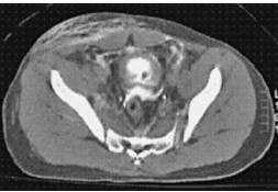
A45-year-old man was kicked several times in the abdomen in a bar fight. He came to the ED and noted that he has not voided for 24 hours. Insertion of a Foley catheter revealed gross hematuria, which persisted after irrigation. A CT scan of the abdomen and pelvis is obtained that does not show any evidence of renal laceration. ACT cystogram is then obtained and is shown in Figure. Appropriate management of this injury includes which of the following?
A. urinary catheter drainage
B. urinary catheter drainage with continuous bladder irrigation
C. bilateral nephrostomy tubes
D. exploratory laparotomy with oversewing of the bladder wall
E. observation
Correct Answer: D Section: (none)
Explanation: Bladder ruptures are highly associated with pelvic fractures. They typically present with hematuria and are typically evaluated with CT cystogram in the setting of trauma. The management of the injury is defined by the location of the rupture. If the rupture remains contained in the extraperitoneal space the treatment is Foley catheter drainage, which allows the bladder to heal spontaneously. However, if the patient has an open pelvic fracture or has other intraabdominal injuries requiring operative exploration, the bladder injury should also be repaired. If imaging of the urinary tract demonstrates rupture of the bladder contents into the peritoneal cavity, operative exploration with a two-layer closure of the defect is the standard of care. Asuprapubic catheter is then placed to help protect the repair.
Question 498:
A 54-year-old male presents to the ED with acute onset of severe abdominal pain. His history is significant for gnawing epigastric pain that radiates to the back for several months. Physical examination demonstrates mild hypertension and tachycardia as well as a rigid "board like" abdomen with generalized rebound tenderness and hypoactive bowel sounds. Rectal examination reveals dark hemoccult positive stools without gross blood. Which of the following would be the next appropriate step in management?
A. order upright chest and abdomen x-rays
B. obtain a CT scan of the abdomen and pelvis
C. plan for upper GI endoscopy
D. take patient to the OR for immediate exploratory laparotomy
E. schedule the patient to be seen in surgery clinic in 1 week
Correct Answer: A Section: (none)
Explanation:
The patient's history of gnawing epigastric pain is consistent with ulcer disease. His presentation is that of a perforated duodenal ulcer. The most appropriate first step is to obtain upright plain films of the chest and abdomen to look for free intraperitoneal air. Although the patient is in mild distress, he is not toxic and it is reasonable to confirm your suspicion with radiologic studies. If the plain films did not demonstrate free air and the patient remained hemodynamically stable, a CT scan of the abdomen and pelvis may be indicated to try to make the diagnosis. However, if the patient did show signs of increasing toxicity and evidence for sepsis, such as hypotension or mental status changes, it would be reasonable to proceed with an exploratory laparotomy to make the diagnosis. Upper endoscopy is not indicated in the acute management of a perforated duodenal ulcer and this patient is currently in significant distress and discharging to home with delayed follow-up is unwise. The patient most likely has a posterior perforation of a duodenal ulcer that has eroded into the gastroduodenal artery causing bleeding per rectum, tachycardia, and hypotension. Diverticulosis is a common cause of bright red blood per rectum in elderly patients but is often painless and not consistent with the presentation of this patient. A ruptured AAA generally presents with hypotension and profound shock. A distended abdomen and pulsatile mass can be found on physical examination. Ruptured esophageal varices present with upper GI bleeding and hematemasis and are most often associated with patients who have chronic liver disease.
Question 499:
A 24-year-old male is involved in a house fire. His sputum is carbonaceous and he has suffered second-and third-degree burns to 65% of his total body surface area (TBSA). He is intubated in the ED without difficulty. A capnometer is placed at the end of the endotracheal tube and there is positive change in color. Examination of his chest reveals bilateral equal breath sounds. Suddenly he experiences ECG changes and goes into cardiac arrest. Which of the following drugs is most likely to be responsible for this event?
A. etomidate
B. rocuronium
C. succinylcholine
D. midazolam
E. ketamine
Correct Answer: C Section: (none)
Explanation:
Succinylcholine is a depolarizing neuromuscular blocking agent that can cause arrhythmias including bradycardia and junctional rhythms because of vasotonic effects. Additionally, succinylcholine is associated with a transient hyperkalemia that can be profound in patients with burns or those who have experienced significant crush injury and result in cardiac arrest.
Question 500:
Which anatomic location is the most common site of extra-adrenal pheochromocytomas?
A. duodenum
B. inferior pole of the kidney
C. paraaortic area
D. parasplenic area
E. peripancreatic area
Correct Answer: C Section: (none)
Explanation:
Pheochromocytomas arise from neuroectoderamal cells and are most often found within the adrenal medulla. Ten percent of all pheochromocytomas are located outside of the adrenal gland along the embryologic path of the adrenal gland. The most common site of an extra-adrenal pheochromocytoma is in the paraaortic area.
Nowadays, the certification exams become more and more important and required by more and more enterprises when applying for a job. But how to prepare for the exam effectively? How to prepare for the exam in a short time with less efforts? How to get a ideal result and how to find the most reliable resources? Here on Vcedump.com, you will find all the answers. Vcedump.com provide not only USMLE exam questions, answers and explanations but also complete assistance on your exam preparation and certification application. If you are confused on your USMLE-STEP-3 exam preparations and USMLE certification application, do not hesitate to visit our Vcedump.com to find your solutions here.