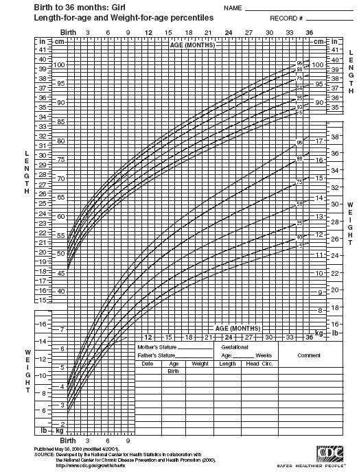
A mother brings her baby girl for a 9-month wellchild visit. You have been following her since birth. Her growth chart is shown in Figure. Her vital signs and examination are otherwise normal.
Correct Answer: A Section: (none)
Explanation:
This infant's growth pattern is most consistent with nutritional FTT. This is often termed “nonorganic” FTT. This term is used for conditions in which the child, usually an infant, begins to fall off of the standardized growth curves. The growth curve in this vignette shows that this infant's weight has trailed off while her length has remained stable. Causes of poor growth that are hormonal in nature will tend to have blunted growth velocity (decreased linear growth) that results in infants and children with short stature and normal weight. Short stature refers to deceased linear growth (i.e., length or height). The infant in this vignette does not have short stature, as her linear growth is normal. Achild with GH deficiency would be expected to have a decreased linear growth velocity (height) with a weight that remains relatively stable. The next best step in the evaluation of this infant would hinge on understanding the total calories that this infant is consuming. A measure of the appropriate caloric intake is related in terms of calories per kilogram per day. This will give a metric to measure whether infants are getting appropriate nutritional intake. Obtaining a serum GH level is an unreliable way to look at an infant's growth due to its pulsatile nature. While a serum somatomedin-C (ILGF-1) may be a more accurate measure of GH activity, in this child a level will likely not reveal much useful information. In children with malnutrition or caloric deprivation, a somatomedin-C level may be depressed due to decreased body mass. If there were a family history of short stature, that would be manifested with poor linear growth, which is not the case in this vignette. While a malabsorption may be a cause of FTT, an UGI series would not be the modality to evaluate for it.
Question 72:
A 29-year-old woman complains of fatigue and decreased exercise tolerance. She takes no medications
and denies changes in the color of the stool. Physical examination is significant for pale skin and
conjunctivae. Stool was negative for blood. Laboratory evaluation revealed Hgb of 7.8 g/dL,
reticulocytopenia, microcytosis, and hypochromia.
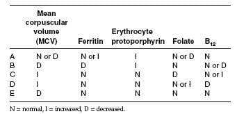
Which of the following would most likely be found on further laboratory testing?
Correct Answer: B Section: (none)
Explanation: Iron-deficiency anemia (IDA) is characterized by a low MCV, low ferritin, and a high erythrocyte protoporphyrin in serum. Microcytosis and hypochromia are the hallmark in the peripheral smear. Elevated erythrocyte protoporphyrin in serum can also be seen in anemia of chronic disease and chronic lead poisoning. The USPSTF recommends screening pregnant women for IDA, but found insufficient evidence to recommend for or against routine screening in other asymptomatic persons. However, the guidelines did recommend routine iron supplementation in asymptomatic infants 6–12 months of age who are at high risk of IDA. Infants are considered to be at high risk if they are living in poverty; are Black, Native American, or Alaskan Native; are immigrants from a developing country; are preterm or low birth weight; or if their primary dietary intake is unfortified cow's milk. The most common cause of cobalamin deficiency is pernicious anemia. Rarely, hypersecretion of gastric acid (i.e., Zollinger-Ellison syndrome) results in cobalamin deficiency. The peripheral smears in folate and cobalamin deficiency are indistinguishable, showing macrocytosis and hypersegmented neutrophils. Both methylmalonic acid and homocysteine levels become elevated with cobalamin deficiency. Folate deficiency is caused by decreased intake, increased utilization, or impaired absorption. Because body stores of folate are low, persons who have an inadequate consumption will become anemic in several months. The recommended amount of dietary folate is 400 µg/day. Anemia is not a diagnosis in itself; it is an objective sign of the presence of a disease. It is always secondary to an underlying condition. In most cases, a thorough history and physical examination can help elicit the pathogenesis of the anemia and direct appropriate treatment.
Question 73:
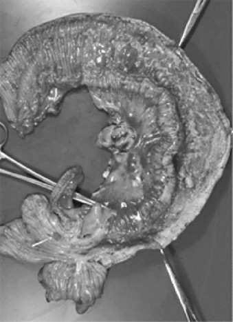
The specimen shown in Figure below, was removed during an exploratory laparotomy of a 22-year-old male who went to surgery because of an intestinal obstruction. What is the most likely diagnosis for the lesion shown in this image?
A. intestinal infarction
B. ulcerative colitis
C. Crohn's disease
D. intestinal tuberculosis
E. small bowel carcinoma
Correct Answer: C Section: (none)
Explanation:
This image shows the typical segmental involvement of the terminal ileum seen in Crohn's disease. There
is no evidence for hemorrhagic infarction. Ulcerative colitis may involve the terminal ileum, but is less likely
as are intestinal tuberculosis and small bowel carcinoma.
Question 74:
A 20-year-old man undergoes a colonoscopy for abdominal pain, weight loss, and diarrhea. Pathologic evaluation reveals transmural chronic inflammation with often noncaseating granulomas. What is the most likely diagnosis?
A. ulcerative colitis
B. ischemic colitis
C. pseudomembranous colitis
D. Crohn's disease E. celiac sprue
Correct Answer: D Section: (none)
Explanation:
Crohn's disease is a chronic inflammatory disorder of unknown etiology that has the potential to involve the different portions of the gastrointestinal (GI) tract from the mouth to the anus. It is characterized by a transmural inflammation with skipped areas in which the intestinal wall is not affected. Frequently, the presence of noncaseating epithelioid granulomas is seen.
Question 75:
A 28-year-old man presents with vague complaints of fatigue, nausea, and dyspnea. He has had occasional episodes of hemoptysis and some burning with urination. Laboratory studies reveal a mild anemia and an elevation of both blood urea nitrogen (BUN) and creatinine. Urinalysis reveals the presence of blood, protein, and red cell casts. Antibodies against which of the following is the cause of this condition?
A. platelet surface antigens
B. basement membrane
C. parietal cell antigens
D. colonic mucosal cells
E. macrophage receptors
Correct Answer: B Section: (none)
Explanation:
Goodpasture syndrome is characterized by antibasement antibodies, with the lungs and kidneys bearing the brunt of the damage. Antibodies against platelet surface antigens, parietal cells, antigens, receptors, and colonic mucosal cells are seen in autoimmune thrombocytopenia, pernicious anemia, myasthenia gravis, and ulcerative colitis, respectively.
Question 76:
A49-year-old female noticed that, in the morning, the small joints of her hands are swollen, painful, and
stiff. Her rheumatoid factor is reportedly strongly positive. Citruline tests (cyclic citrullinated peptide [CCP])
are also positive.
What disease does the patient most likely have?
A. degenerative joint disease
B. rheumatoid arthritis
C. spondyloarthritis
D. tennis elbow
E. septic arthritis
Correct Answer: B Section: (none)
Explanation:
In this case, the patient most likely has rheumatoid arthritis, an autoimmune chronic relapsing disorder that mostly affects the joints. The disease is usually seen in Western European and North American White females between the ages of 30 and 50. The clinical hallmark of the disease is symmetric swelling of the small joints of the hands and feet, particularly the proximal and interphalangeal joints. Swelling, pain, and stiffness are most severe in the morning. Pathologically, a pannus, or hypertrophic inflamed synovium, is produced that may eventually erode into the articular cartilage, with subsequent fibrosis, restriction of movements, and deformity.
Question 77:
Which of the following patients is most likely to progress to develop cirrhosis?
A. a man with chronic liver disease due to cytomegalovirus infection
B. a man with chronic liver disease due to hepatitis B
C. a man with chronic liver disease due to hepatitis C
D. a man chronic with liver disease due to hepatitis D
E. a man with chronic liver disease due to human immunodeficiency virus
Correct Answer: C Section: (none)
Explanation:
Hepatitis C which is caused by a singlestranded RNA virus is responsible for over 90% of the cases of hepatitis associated with transfusion of blood and blood products in the United States. The disease also occurs in drug abusers and transplant recipients, as well as renal dialysis patients. It is associated with a higher incidence of chronic hepatitis, which occurs in 50% of those affected and cirrhosis complicates 20% of the cases.
Question 78:
Which factor is most directly related to prognosis in a patient with the diagnosis of squamous cell carcinoma of the esophagus?
A. degree of differentiation
B. duration of the symptoms
C. method of treatment
D. stage at the time of diagnosis
E. type of symptoms
Correct Answer: D Section: (none)
Explanation:
Carcinoma of the esophagus is a highly lethal tumor and is a disease of the elderly. Etiology factors include alcoholism, cigarette smoking, hot drinks, aflatoxins, and smoked fish. The overall prognosis is very poor with 70% of the patients dying within 1 year after diagnosis of the disease. The most important parameter of the prognosis is the stage at the time of diagnosis, because over 80% 5-year survival is present in the tumors detected during the surveillance of Barrett esophagus.
Question 79:
An 18-year-old male developed chills, fever, and a painful swollen knee. What test would be most appropriate in order to help in making the diagnosis?
A. culture of joint fluid from the affected knee
B. Lyme disease test
C. MRI
D. serum protein electrophoresis
E. study of crystals in the synovium
Correct Answer: A Section: (none)
Explanation:
Because we suspect that this patient has suppurative arthritis, the test to be ordered would be a culture of the joint fluid from the affected knee to ascertain which organism is involved and, with further identification and sensitivity, to determine which would be the antibiotic of choice. It would also be important to determine whether this is a hematogenous spread, secondary to osteomyelitis or contamination of the joint by a wound.
Question 80:
A 28-year-old male undergoes an orchiectomy because of a suspicious testicular mass. The pathologic evaluation reveals the tumor to have gross and microscopic hemorrhage and necrosis. The tumor is noted to be composed mostly of cytotrophoblastic and syncytiotrophoblastic cells. What is this patient's diagnosis?
A. embryonal carcinoma
B. seminoma
C. teratoma
D. choriocarcinoma
E. yolk sac tumor
Correct Answer: D Section: (none)
Explanation:
Choriocarcinoma is a rare tumor of the testicles, but it is characterized by hemorrhage and necrosis. This tumor comprises only 1% of the malignant germ cell tumors and is rarely seen in a pure form. In general, they are small (no larger than 5 cm in diameter) and human chorionic gonadotropin (hCG) can be readily demonstrated in the blood. The cells seen in the hemorrhagic areas are cytotrophoblastic, as well as syncytiotrophoblastic, cells.
Nowadays, the certification exams become more and more important and required by more and more enterprises when applying for a job. But how to prepare for the exam effectively? How to prepare for the exam in a short time with less efforts? How to get a ideal result and how to find the most reliable resources? Here on Vcedump.com, you will find all the answers. Vcedump.com provide not only USMLE exam questions, answers and explanations but also complete assistance on your exam preparation and certification application. If you are confused on your USMLE-STEP-3 exam preparations and USMLE certification application, do not hesitate to visit our Vcedump.com to find your solutions here.