A 60-year-old male developed painless hematuria. On further clinical evaluation, a CT scan showed a 7 cm mass on the lower pole of the right kidney. What would be your most likely diagnosis in this case?
A. neuroblastoma
B. medullary fibroma
C. Wilms tumor
D. transitional cell carcinoma
E. renal cell carcinoma
Correct Answer: E Section: (none)
Explanation: The most important and frequent cause of painless hematuria is renal cell carcinoma. This symptom is usually associated with a palpable mass on the flank, as well as costovertebral pain. Occasionally, renal cell carcinomas are associated with a paraneoplastic syndrome, which includes polycythemia, hypercalcemia, hypertension, feminization or masculinization, Cushing syndrome, and so on. The other answers listed are mostly seen in children. Transitional cell carcinoma is rarer than renal cell carcinoma and medullary fibroma is a benign tumor.
Question 82:
Asmall area of abnormality is noted on the lateral wall of the urinary bladder during a cystoscopy on a patient being evaluated for asymptomatic microscopic hematuria. Biopsy of the lesion is most likely to reveal which of the following?
A. adenocarcinoma
B. neuroendocrine carcinoma
C. rhabdomyosarcoma
D. squamous cell carcinoma
E. papillary transitional cell carcinoma
Correct Answer: E Section: (none)
Explanation:
Papillary cancer arises most frequently from the lateral and posterior bladder walls. At cystoscopy, tumors may be small, delicate, lowgrade papillary lesions limited to the mucosal surface or larger high grade, solid, and invasive which are often ulcerated. Papillary and exophitic cancers tend to be better differentiated. Infiltrating tumors are usually more anaplastic. Nonurothelial forms of bladder cancer are squamous cell carcinomas, adenocarcinomas, neuroendocrine carcinomas, and rhabdomyosarcomas. The frequency of these tumors is much lower
Question 83:
Which of the following is the most common characteristic of a serous cystadenocarcinoma of the ovary?
A. It causes pseudomyxoma peritonei.
B. It is composed of transitional epithelial cells.
C. It is frequently bilateral.
D. It is most common in children and young adults.
E. It often metastasizes to the brain.
Correct Answer: C Section: (none)
Explanation:
Serous cystadenocarcinoma of the ovary is the most common malignant ovarian tumor and is frequently bilateral. Microscopically, they show a variegated appearance with papillary pattern. Different degrees of anaplasia of the cuboidal to columnar cells cover the papilla and occasional calcified concretions (Psammoma bodies) are present. These tumors almost never metastasize to the brain and they are not seen in children or young adults.
Question 84:
Ahistory of which of the following conditions would result in the greatest increase in the likelihood of developing colon cancer?
A. Crohn's disease
B. diverticulosis
C. hamartomatous polyp
D. pseudomembranous colitis
E. ulcerative colitis
Correct Answer: E Section: (none)
Explanation:
Adenocarcinoma of the colon is the most common type of malignancy arising in the large intestine. Iron deficiency and microcytic anemia may be the presenting symptoms due to the bleeding from the tumor's ulceration. Alternatively, the tumor may be suspected by detention of occult fecal blood test, bowel obstruction, or through the development of hepatic enlargement secondary to metastasis. The gross appearance of this tumor is usually polypoid and ulcerated. Many ulcerating tumors involve the full circumference of the bowel and appear radiologically as an "apple core" lesion. The microscopic appearance is that of gland-forming malignant cells and usually mucin production is present. The prognosis is related to the stage of the disease. Ulcerative colitis is an inflammatory disease of uncertain etiology that has a relapsing course. Patients with ulcerative colitis have a higher than normal incidence of developing colon carcinoma, approximately 10%. Carcinoid tumors originate in the neuroendocrine cells throughout the intestinal tract. The appendix is most frequently involved, followed by the terminal ileum. On histologic examination, carcinoids are composed of uniform, round cells forming small nests or cords without encapsulation. Special stains performed show neurosecretory granules in the cytoplasm, which are positive for chromogranin, neuron-specific enolase, and other staining.
Question 85:
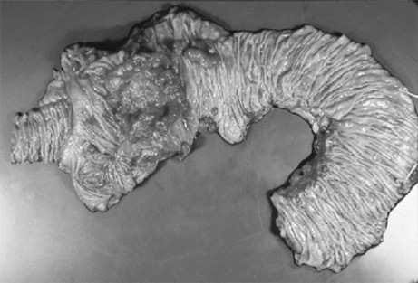
A 65-year-old postmenopausal woman relates a complaint of being excessively tired for 6 months. Her laboratory results were remarkable for a microcytic anemia. A colonoscopy followed by a biopsy revealed a mass of the right colon. After the initial biopsy, a right colectomy was performed shown in figure below. What would be the most likely diagnosis?
A. adenomatous polyp
B. lipoma of the cecal valve
C. adenocarcinoma of the right colon
D. ischemic colitis
E. Crohn's disease
Correct Answer: C Section: (none)
Explanation: Adenocarcinoma of the colon is the most common type of malignancy arising in the large intestine. Iron deficiency and microcytic anemia may be the presenting symptoms due to the bleeding from the tumor's ulceration. Alternatively, the tumor may be suspected by detention of occult fecal blood test, bowel obstruction, or through the development of hepatic enlargement secondary to metastasis. The gross appearance of this tumor is usually polypoid and ulcerated. Many ulcerating tumors involve the full circumference of the bowel and appear radiologically as an "apple core" lesion. The microscopic appearance is that of gland-forming malignant cells and usually mucin production is present. The prognosis is related to the stage of the disease. Ulcerative colitis is an inflammatory disease of uncertain etiology that has a relapsing course. Patients with ulcerative colitis have a higher than normal incidence of developing colon carcinoma, approximately 10%. Carcinoid tumors originate in the neuroendocrine cells throughout the intestinal tract. The appendix is most frequently involved, followed by the terminal ileum. On histologic examination, carcinoids are composed of uniform, round cells forming small nests or cords without encapsulation. Special stains performed show neurosecretory granules in the cytoplasm, which are positive for chromogranin, neuron-specific enolase, and other staining.
Question 86:
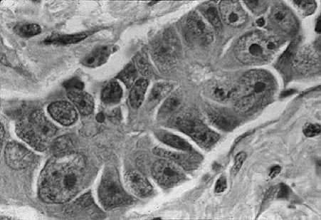
A62-year-old male, who has been smoking two packs of cigarettes a day for the past 35 years, was found to have a 2.5 cm peripheral solitary nodule in the left upper lobe of lung. Thoracotomy with biopsy was performed and a picture of the biopsy findings is depicted in Figure below. With the clinical information and the biopsy findings what would be the most likely diagnosis?
A. pulmonary infarct
B. adenocarcinoma
C. small cell anaplastic carcinoma
D. tuberculosis
E. granulomatous inflammation.
Correct Answer: B Section: (none)
Explanation:
The incidence of adenocarcinoma of the lung has increased significantly in the last two decades and is now the most common form of lung cancer in women and, in some studies, also in men. There may be mixtures of histologic patterns in the same cancers and, therefore, the finding of squamous cell carcinoma is not infrequent. A recent classification that is more common for clinical use has been developed in response to the necessity for the different therapies. These two large groups are divided into small cell versus nonsmall cell carcinomas. On histologic examination, the adenocarcinomas can be divided into bronchial-derived adenocarcinoma and bronchioloalveolar carcinoma. This classification is based on histologic findings alone. The lesions, in general, are peripherally located and tend to be smaller. Adenocarcinoma, including the bronchioloalveolar variant, is the least frequently associated with a history of cigarette smoking. Special stains for mucin are frequently positive.
Question 87:
Of the following, which is the best indicator of response to treatment or progression of disease in monitoring patients being treated for prostate cancer?
A. digital rectal examination
B. serum prostate-specific antigen (PSA)
C. computed tomography (CT) scans
D. MRI
E. positron emission tomography (PET) scans
Correct Answer: B Section: (none)
Explanation:
At present, we have widespread screening programs for prostatic cancer using the digital rectal examination in combination with the serum PSA. These detect most of the malignant processes in the prostate. Patients with elevated serum PSA are further evaluated with needle biopsies. Preoperative PSA levels are correlated with the cancer volume. Serum PSA levels are a useful monitor for response to
treatment and progression or recurrence of disease following therapy.
Question 88:
A54-year-old woman has a dilation and curettage procedure for the evaluation of postmenopausal bleeding. Which of the following pathologic diagnoses would carry the most favorable prognosis for the patient?
A. well-differentiated adenocarcinoma with a squamous differentiation
B. serous carcinoma
C. clear cell adenocarcinoma
D. carcinosarcoma
E. carcinosarcoma with heterologous elements
Correct Answer: A Section: (none)
Explanation:
One-third of all endometrial adenocarcinomas contain squamous cells, in addition to glandular elements. If the squamous element is well differentiated with no more than minimal atypia, the tumor is called well-differentiated adenocarcinoma with squamous differentiation. These tumors enjoy a better prognosis stageby-stage compared with all of the others listed in the question. These tumors are less common and they show an aggressive behavior. The histologic grading, therefore, is not of clinical value. These aggressive tumors are serous carcinoma, clear cell adenocarcinoma, carcinosarcoma.
Question 89:
A 17-year-old male is evaluated for a painless neck mass. You assess the mass as lymphadenopathy and arrange for a biopsy. The pathology report subsequently notes the presence of Reed-Sternberg cells. Which of the following is the most likely diagnosis?
A. Hodgkin lymphoma
B. non-Hodgkin lymphoma
C. metastatic testicular cancer
D. acute lymphocytic leukemia
E. papillary carcinoma of the thyroid
Correct Answer: A Section: (none)
Explanation:
The Reed-Sternberg cell can be classified as the classic type, the mononuclear variant, the lymphocytic histiocytic variant, lacunar and pleomorphic variant. The classic Reed-Sternberg cell is a binucleated cell that contains an ovoid-shaped nucleus with regular contours and prominent eosinophilic nucleoli. Cytoplasm is abundant and eosinophilic. On cytogenetic studies, the Reed-Sternberg cells are either aneuploid or frequently hypertetraploid. The classic Reed-Sternberg cell is thought to be an end-stage cell that does not divide. The mononuclear variants of the Reed-Sternberg cells (so-called Hodgkin cells) could be identified in any type of Hodgkin disease, but they are not diagnostic of Hodgkin's.
Question 90:
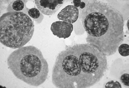
A 67-year-old female was admitted to the hospital because of chronic fatigue and low back pain. An x-ray of the vertebral column showed diffuse osteoporosis and compression fractures of L1 and L2 vertebral bodies. The complete blood count (CBC) was within normal limits. The peripheral blood smear showed rouleaux formation. The immunoelectrophoresis showed a monoclonal spike of more than 3 g. A bone marrow biopsy was performed and showed an increase of more than 20% in plasma cells see Figure below Microscopically, the bone marrow examination will reveal which of the following?
A. normocellular marrow with normal hematopoiesis
B. an increase in myeloid elements
C. increase in megakaryocytes
D. increase in mature lymphocytes
E. increase in plasma cells, usually more than 30% of the total cells
Correct Answer: E Section: (none)
Explanation:
Multiple myeloma is a plasma cell dyscrasia that is characterized by involvement of the skeleton in multiple sites. The characteristic x-ray shows punched-out bone lesions that are very easily seen in the calvarium. Extension of the disease to lymph nodes and extranodal sites, such as skin, can be seen. The bone marrow biopsy and smears reveal an increased number of plasma cells, which usually constitute greater than 20% of all of the cells. The cells either diffusely infiltrate and replace the marrow elements or can be seen scattered throughout the hematopoietic elements. The neoplastic plasma cells have a perinuclear hof and an eccentrically placed nucleus which allows the recognition. In 99% of patients with multiple myeloma, electrophoretic analysis reveals increased levels of IgG in the blood, light chains (Bence-Jones proteins) in the urine, or both. The monoclonal IgG produces a high spike when seen in the serum or in the urine, subject to electrophoresis. In general, the quantitative analysis of the monoclonal IgG is more than 3 g. The clinicopathologic diagnosis of multiple myeloma rests on radiographic and laboratory findings. Marrow examination may reveal increased plasma cells or sheet-like aggregates that may completely replace the normal elements. The prognosis for this condition is variable, but generally poor.
Nowadays, the certification exams become more and more important and required by more and more enterprises when applying for a job. But how to prepare for the exam effectively? How to prepare for the exam in a short time with less efforts? How to get a ideal result and how to find the most reliable resources? Here on Vcedump.com, you will find all the answers. Vcedump.com provide not only USMLE exam questions, answers and explanations but also complete assistance on your exam preparation and certification application. If you are confused on your USMLE-STEP-3 exam preparations and USMLE certification application, do not hesitate to visit our Vcedump.com to find your solutions here.