A 79-year-old woman presents with a complaint of chronic and progressive mid-back pain for several months but no other symptoms. Spinal x-rays reveal two compression fractures of the T10 vertebral body. Calcium, phosphate, and alkaline phosphatase levels are all within normal range. Histological examination of the affected vertebral bone would most likely reveal which of the following?
A. bony necrosis with acute and chronic inflammation and granulomas
B. disorganized interconnecting trabeculae of woven bone rimmed by osteoblasts
C. thickened, irregular trabeculae with prominent cement lines
D. thinning of the cortical and trabecular bone
E. widened osteoid seams of peripheral trabeculae
Correct Answer: E
Section: Pathology and Path physiology This woman is suffering from senile, or postmenopausal, osteoporosis. In such patients, the calcium and phosphate levels will typically be within the reference range. The alkaline phosphatase will also usually be normal, but may be increased following fractures. Microscopic examination of affected bones will reveal loss of bone mass tending to affect horizontal more than vertical trabeculae. Bony necrosis with acute and chronic inflammation and granulomas (choice A) occurs in tuberculosis infection of the spine, where it is referred to as Pott disease. Disorganized interconnecting trabeculae of woven bone rimmed by osteoblasts (choice B) describes the appearance of an osteoblastoma, a benign bone tumor that is histologically very similar to and often described in conjunction with an osteoid osteoma. A characteristic difference between these two benign bone tumors is that osteoid osteomas are very painful lesions (excess prostaglandin E production) that respond dramatically to aspirin, while osteoblastomas produce a dull, achy pain that does not respond to aspirin. Thickened, irregular trabeculae with prominent cement lines (choice C) describe the morphology seen in Paget disease. Widened osteoid seams of peripheral trabeculae (choice E) reflect the appearance of unmineralized osteoid found in osteomalacia. Alkaline phosphatase levels are typically increased in osteomalacia (and rickets) and Paget disease.
Question 212:
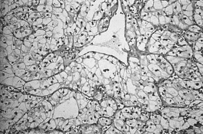
A 64-year-old man presents to his family physician with hematuria and flank pain. A radiology study identifies a renal mass. Aphotograph of this renal lesion's histology is displayed in below figure. The kidney mass is most likely which of the following?
A. angiomyolipoma
B. oncocytoma
C. renal cell carcinoma
D. transitional cell carcinoma
E. xanthogranulomatous pyelonephritis
Correct Answer: C
Section: Pathology and Path physiology Figure shows a clear cell adenocarcinoma, which is the most common histologic appearance for renal cell carcinoma. The patient's age, gender, and clinical presentation are typical for this malignant neoplasm. Angiomyolipoma (choice A) is a benign renal tumor that displays a mixture of blood vessels, smooth muscle, and mature fat on microscopic examination. Oncocytoma (choice B) is a benign renal neoplasm constructed by monomorphic cells with granular eosinophilic cytoplasm. Transitional cell carcinoma (choice D) usually arises in the renal pelvis and histologically is composed of anaplastic transitional cells without a clear cell adenocarcinoma component. Xanthogranulomatous pyelonephritis (choice E) is a benign inflammatory condition of the kidney that may produce a mass effect. The gross appearance, but not the microscopic appearance, may be confused with renal cell carcinoma.
Question 213:
A 14-year-old, severely physically disabled individual is now on a respirator. His first 4 years of life were medically uneventful. Over the last 10 years, he has suffered from increasing symmetric muscle weakness that first affected the pelvic girdle and now involves almost all muscle groups. Several years ago, the calf portion of his legs appeared enlarged and on biopsy demonstrated fatty pseudohypertrophy with random alternating muscle fiber atrophy and hypertrophy. Which of the following is the most likely diagnosis?
A. cerebral palsy
B. muscular dystrophy
C. myositis ossificans
D. poliomyelitis
E. trichinosis
Correct Answer: B
Section: Pathology and Path physiology The clinical scenario describes a classic example of Duchenne muscular dystrophy. The weakness is symmetric and most often begins at the pelvic girdle. Fatty pseudohypertrophy with alternating muscle fiber atrophy and hypertrophy typify the histologic changes. Many patients die before reaching their 20s. The other diagnostic options, cerebral palsy (choice A), myositis ossificans (choice C), poliomyelitis (choice D), and trichinosis (choice E), do not fit the clinical picture.
Question 214:
A 48-year-old man with a long history of chronic viral hepatitis secondary to chronic hepatitis B virus (HBV) infection presented with a 2-month history of weight loss. Physical exam showed a cachectic man in mild distress. Scleral icterus and abdominal distension were present. Workup showed the presence of two mass lesions within the liver on ultrasound, which were confirmed on CT scan. Laboratory values revealed the following: AST 161 U/L; ALT U/L 188; total bilirubin 6.8 mg/dL; direct bilirubin 4.9 mg/dL; alkaline phosphatase 245 U/L; total protein 5.9 g/dL; albumin 2.8 g/dL; HBsAg positive; anti-HBs negative; anti-HCV negative; alpha-fetoprotein 7200 ng/mL. Aneedle biopsy of one of the mass lesions was performed under CT guidance. Which of the following is the most likely diagnosis?
A. fholangiocarcinoma
B. focal nodular hyperplasia
C. hepatocellular carcinoma
D. liver cell adenoma
E. metastatic carcinoma
Correct Answer: C
Section: Pathology and Path physiology One of the potential consequences of chronic hepatitis B (and C) infection is the development of hepatocellular carcinoma, and the presence of the mass lesions with high levels of alphafetoprotein makes this extremely likely in this case and will be confirmed by the needle biopsy. Cholangiocarcinoma (choice A) is unusual in the United States but has a much higher incidence in Asia in association with infestation by the liver fluke, C. sinensis. Focal nodular hyperplasia (choice B) is a mass lesion of the liver, but is not a true neoplasm. It has a characteristic central scar and microscopically resembles cirrhosis. Liver cell adenoma (choice D) typically occurs in women taking oral contraceptives. Metastatic carcinoma (choice E) is certainly a possibility but given the clinical history of this case plus the high alpha-fetoprotein and the lack of any evidence for a primary malignant tumor elsewhere, hepatocellular carcinoma remains the most probable diagnosis.
Question 215:
A 57-year-old man has just returned from an overseas trip and reports having had severe substernal chest pain 3 days ago. Which of the following is the most appropriate laboratory test to order for this patient?
A. aspartate aminotransferase
B. creatine kinase, MB fraction
C. creatine kinase, total
D. lactate dehydrogenase, LD1 fraction
E. lactate dehydrogenase, total
F. troponin I
Correct Answer: F
Section: Pathology and Path physiology Troponin I is now the method of choice for the laboratory diagnosis of MI. There is a detectable increase within 48 hours of the infarction and the peak level is reached within 1436 hours. Levels do not return to baseline for 310 days making it an appropriate test for this patient who was 3 days post- infarction. Aspartate aminotransferase (choice A) was the first serum enzyme marker used for the diagnosis of MI, but it has poor specificity and sensitivity compared to newer markers and is no longer used for this purpose. Creatine kinase, total (choice C) is not used for the diagnosis of MI since the cardiac fraction (MB) can be overwhelmed by the presence of the skeletal muscle fraction. Creatine kinase-MB (choice B) is still being used in some institutions but it returns to baseline in 23 days and would not be useful for this patient. Lactate dehydrogenase, either LD1 fraction (choice D) or lactate dehydrogenase, total (choice E) return to baseline later than creatine kinase but have been replaced by troponin I and are seldom used.
Question 216:
Parents of an 18-month-old boy bring their child to their pediatrician as they are concerned about their son's recurring skin and lung infections. Biopsy of one of the boy's current skin lesions reveals the presence of neutrophils, lymphocytes, and scattered epithelioid cell granulomas. Culture of the lesion is positive for Staphylococcus aureus. Which of the following would best account for this patient's condition?
A. absence of T-helper cell activity
B. adenosine deaminase enzyme deficiency
C. complement C3b opsonization abnormality
D. decreased hydrogen peroxide production
E. defective macrophage phagocytosis
Correct Answer: D
Question 217:
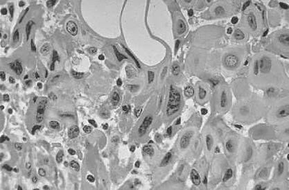
Aretroperitoneal mass was discovered during exploratory surgery on a 49-year-old woman. A photomicrograph of a section taken from the mass is shown in below figure. Which of the following terms would be most appropriate to describe its appearance?
A. anaplasia
B. aplasia
C. dysplasia
D. hyperplasia
E. metaplasia
Correct Answer: A
Section: Pathology and Path physiology The cells in figure show dramatic differences in the size, shape, and staining intensity of nuclei as well as differences in the cells overall, which allows one to say that they are pleomorphic. An additional factor is the lack of differentiation of these cells such that the cell type cannot be recognized, which is the definition of anaplasia. Aplasia (choice B) is a lack of growth of the anlage or primordium of an organ. Dysplasia (choice C) at the cellular level refers to disorderly maturation, usually of an epithelium. Hyperplasia (choice D) is an increase in the number of cells and this process may be either physiological or pathological. Metaplasia (choice E) is the replacement of one mature cell type by another (e.g., columnar epithelium being replaced by squamous epithelium in a smoker's lung).
Question 218:
A 37-year-old man visiting a third world country drinks from a fecally contaminated source of water. During the next week he has gradually increasing fever, anorexia, myalgia, and headache. He subsequently develops a maculopapular rash on his abdomen and his fever increases to 104°F with abdominal pain and splenomegaly. During the third week, his condition rapidly deteriorates with intestinal bleeding, shock, and death. An autopsy reveals ulcerations overlying the Peyer patches of the small intestine, one of which is perforated. Which of the following is the most likely diagnosis?
A. amebiasis
B. cholera
C. cryptosporidiosis
D. giardiasis
E. typhoid fever
Correct Answer: E
Section: Pathology and Path physiology This is a case of typhoid fever. During the prodromal stage there is a gradual, step-like increase in fever with malaise, anorexia, myalgia, and headache. By the second week, the fever usually plateaus and the patient is very sick. There may be constipation or diarrhea with abdominal pain and distention, weakness, and a maculopapular rash (rose spots), particularly on the abdomen. If there are no complications, the patient may gradually improve over the next 2 weeks. One of the classic pathologic findings in typhoid is intestinal ulcerations over hyperplastic and necrotic Peyer patches. These may perforate and hemorrhage, as in this patient. Amebiasis (choice A) can produce a range of symptoms from being subclinical to producing fulminant dysentery. However, it involves the large bowel rather than the small intestine and produces flask-shaped ulcers separated by areas of normal bowel. Cholera (choice B) does not actually invade the intestinal epithelium and therefore causes insignificant microscopic changes and no ulceration. Cryptosporidiosis (choice C) causes a severe, chronic diarrhea in AIDS patients. The protozoa attach to the brush border of the intestinal epithelial cells but do not cause ulceration. Giardia (choice D) attaches to duodenal epithelial cells, but does not invade those cells and does not cause ulceration.
Question 219:
A 35-year-old apparently healthy man undergoes a medical examination while applying for life insurance. He is not anemic. His hemoglobin electrophoresis is reported as: HbA, 62%; HbS, 35%; HbF, 1%; HbA2, 1%; no variant C, D, G, or H bands detected. The most likely diagnosis is which of the following?
A. sickle-cell disease
B. sickle thalassemia minor
C. sickle trait
D. thalassemia major
E. thalassemia minor
Correct Answer: C
Section: Pathology and Path physiology Individuals with sickle trait are healthy and not anemic. Hemoglobin electrophoresis demonstrates a minor proportion of hemoglobin S and a major proportion of hemoglobin A. Fetal hemoglobin and A2 hemoglobin are usually normal. Sickle trait confers the benefit of protecting erythrocytes from some forms of malarial infection. About 9% of blacks in the United States have sickle trait. In sickle cell disease (choice A), almost all hemoglobin is hemoglobin S. No hemoglobin A is detected and patients have a clinical history of severe anemia. Sickle thalassemia minor (choice B) presents as a chronic microcytic anemia with a major hemoglobin S component and elevated hemoglobin A2. Thalassemia major (choice D) and thalassemia minor (choice E) do not demonstrate hemoglobin S on electrophoresis.
Question 220:
A 29-year-old woman presents with weakness, fatigue, easy bruising, and nosebleeds. Analysis of her blood reveals a reciprocal translocation between chromosomes 22 and 9, and low leukocyte alkaline phosphatase levels. These findings confirm a diagnosis of which of the following?
A. acute lymphoblastic leukemia
B. Burkitt lymphoma
C. chronic myelogenous leukemia
D. follicular lymphoma
E. Hodgkin lymphoma
Correct Answer: C
Section: Pathology and Path physiology Ninety percent of individuals with chronic myelogenous leukemia have an acquired Philadelphia chromosome abnormality consisting of a translocation between chromosomes 22 and 9. The translocation places the proto-oncogene c-abl from chromosome 9 next to the breakpoint cluster region (bcr) on chromosome 22. The unique gene sequence bcr-abl confers a growth advantage with subsequent clonal expansion. Acute lymphoblastic leukemia (choice A) does not have a known reproducible gross chromosomal derangement. Burkitt lymphoma (choice B) is associated with a translocation of the c-myc oncogene from chromosome 8 to chromosome 14. Follicular lymphoma (choice D) is associated with a translocation between chromosomes 14 and 18. Hodgkin lymphoma (choice E) is characterized by the presence of ReedSternberg cells and has a number of subtypes.
Nowadays, the certification exams become more and more important and required by more and more enterprises when applying for a job. But how to prepare for the exam effectively? How to prepare for the exam in a short time with less efforts? How to get a ideal result and how to find the most reliable resources? Here on Vcedump.com, you will find all the answers. Vcedump.com provide not only USMLE exam questions, answers and explanations but also complete assistance on your exam preparation and certification application. If you are confused on your USMLE-STEP-1 exam preparations and USMLE certification application, do not hesitate to visit our Vcedump.com to find your solutions here.