A 47-year-old gardener receives an insect sting while pruning some rose bushes. Within a few minutes the area around the sting is swollen and red. The swelling is mostly the result of which of the following?
A. decreased plasma oncotic pressure
B. increased hydrostatic pressure
C. increased vascular permeability
D. lymphatic obstruction
E. venous obstruction
Correct Answer: C
Section: Pathology and Path physiology Following tissue injury (in this case caused by the insect sting), vasoactive inflammatory mediators originating from both cellular and humoral sources are released at the site of injury. These produce vasodilation of arterioles and increased blood flow producing the redness, and increased vascular permeability of enules allowing the formation of an exudates that produces swelling. All of the other choices can produce edema, but do not feature an increase in vascular permeability (they produce noninflammatory edema). Decreased plasma oncotic pressure (choice A) can result from either excessive loss (e.g., nephrotic syndrome) or decreased synthesis (e.g., cirrhosis, protein malnutrition) of plasma proteins, principally albumin. Increased hydrostatic pressure (choice B) occurs, for example, in heart failure where the pressure builds up behind the failing pump. Lymphatic obstruction (choice D) occurs where there is blockage to the normal lymphatic drainage. This could be due to the growth of an obstructing cancer or to inflammation and fibrosis (e.g., postsurgery, filariasis). Venous obstruction (choice E) leads to increased hydrostatic pressure as the blood backs up behind the obstruction.
Question 242:
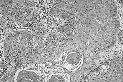
During a routine dental examination of a 55-year-old man, a small, reddish, and raised lesion is noted on the posterior of the median sulcus of his tongue. The patient is referred to a surgeon who performs an excisional biopsy of the lesion. A section of the biopsy is shown in below figure. Which of the following is the most appropriate diagnosis?
A. amyloidosis
B. candidiasis
C. inverted papilloma
D. leukoplakia
E. squamous cell carcinoma
Correct Answer: E
Section: Pathology and Path physiology In figure, the pleomorphism of the nuclei and the clearly invasive nature of these cells indicate the malignant nature of this lesion. Furthermore, three keratin "pearls" at the bottom of the image and the sheets of cells, many with discernable intercellular bridges (prickles), allow one to recognize this as a squamous cell carcinoma. Amyloidosis (choice A) can affect many organs including the tongue. However, amyloid would appear as acellular, eosinophilic deposits around vessels and in the parenchyma of tissues which is not seen here. Candidiasis (choice B) is commonly seen on the tongue, but is not invasive in a person with an intact immune system. Its appearance is that of pseudohyphae with yeast forms. Inverted papilloma (choice C) is a benign but locally aggressive tumor found in the nose and paranasal sinuses but not seen in the tongue. Leukoplakia (choice D) of the oral cavity appears as a white patch that cannot be scraped off and cannot be otherwise identified. This is not the gross description given for this patient.
Question 243:
A 27-year-old woman complains of double vision and drooping of her eyelids. She also states that she has recently noticed weakening of her jaws when chewing tough foods. Physical examination reveals mild weakness of her facial and neck muscles but no atrophy is noted. Which of the following diagnoses is most likely?
A. conjunctivitis
B. myasthenia gravis
C. orbital inflammatory pseudotumor
D. Parkinson's disease
E. polymyositis
Correct Answer: B
Section: Pathology and Path physiology Myasthenia gravis is an autoimmune disease characterized by autoantibodies to acetylcholine receptors, and weakness of both facial and ocular muscles. Conjunctivitis (choice A) defines an inflammatory or infectious condition of the conjunctiva, and is not consistent with this clinical scenario. Orbital inflammatory pseudotumor (choice C) is a benign mass lesion of the eye region and is not associated with muscle weakness and autoantibodies. Parkinson's disease (choice D) is a neurologic movement disorder that does not demonstrate either weakness or acetylcholine autoantibodies. Polymyositis (choice E) is a subacute inflammatory disease of skeletal muscle, typically affecting proximal muscle groups, and specifically does not include facial muscles.
Question 244:
A 67-year-old woman complains of gradually increasing fatigue. On physical examination, she is found to be anemic and has a peripheral neuropathy characterized by loss of position and vibratory sense. Laboratory studies document a macrocytic anemia and decreased WBC and platelets counts. What pathological mechanism accounts for these findings?
A. a diet deficient in folate
B. autoantibodies against parietal cells or intrinsic factor
C. chronic blood loss
D. diabetes mellitus E. myelodysplastic sideroblastic anemia
Correct Answer: B
Section: Pathology and Path physiology The clinical and laboratory findings suggest a diagnosis of pernicious anemia. Almost all cases are due to autoantibodies against parietal cells or intrinsic factor. These autoantibodies disrupt the normal absorption of vitamin B12. The inability to absorb vitamin B12 leads to a macrocytic pancytopenia and peripheral neuropathy. A diet deficient in folate (choice A) can cause a macrocytic anemia, but there are no concomitant neurological findings. Chronic blood loss (choice C) usually results in microcytic hypochromic anemia due to iron deficiency. Anemia and peripheral neuropathy commonly occur with diabetes mellitus (choice D). However, the anemia is normocytic and the neuropathy is sensory. Myelodysplastic sideroblastic anemia (choice E) may present hematologically with a macrocytic pancytopenia. A peripheral neuropathy is not seen.
Question 245:
A 67-year-old man complains of low back pain and generalized weakness, gradually worsening over the past 6 months. Physical examination reveals an individual in moderate discomfort due to the back pain. Laboratory examination reveals an anemia with rouleaux formation of the erythrocytes on the peripheral smear. Urinalysis demonstrates proteinuria and hypercalciuria. X-rays reveal diffuse osteoporosis of the spine and small lytic lesions in the ribs. Which of the following diagnoses most likely explains these findings?
A. fibrous dysplasia
B. iron-deficiency anemia
C. metastatic prostatic carcinoma
D. multiple myeloma
E. osteosarcoma
Correct Answer: D
Section: Pathology and Path physiology This is a patient with multiple myeloma and one of the earliest symptoms of the disease is back pain. These patients have increased levels of Ig in the blood (which produces an increased erythrocyte sedimentation rate and will be seen as rouleaux formation on the blood smear) and light chains (Bence-Jones protein) in the urine. Multiple myeloma causes multifocal osteolytic lesions throughout the skeletal system and these are apparent on x-rays and are also responsible for the hypercalcemia as the ongoing bone destruction releases calcium. Fibrous dysplasia (choice A) is a disorder of bone in children with progressive replacement of a localized area of bone by an abnormal proliferation of benign fibrous tissue and bony trabeculae composed of haphazardly arranged woven bone. It occurs as a monostotic and polyostotic form, but neither could account for the findings in this case. Iron- deficiency anemia (choice B) does not produce any of the findings in this case. Metastatic prostatic carcinoma (choice C) can spread quite easily to the lumbar-sacral spine and this causes back pain. However, these bone lesions are osteoblastic rather than osteolytic. Osteosarcoma (choice E) is typically found in teenagers or young adults. When seen in older individuals, it usually occurs in association with Paget disease of the bone.
Question 246:
Approximately 6 months ago, a 59-year-old man developed a dull, continuous abdominal pain that radiated to the right upper quadrant and was relieved by bending forward. He has also had recurrent thrombophlebitis. He now develops jaundice. Of the following, which is the condition that would most likely explain all of these findings?
A. alcoholic cirrhosis
B. cholecystitis
C. cholelithiasis
D. pancreatic adenocarcinoma
E. viral hepatitis
Correct Answer: D
Section: Pathology and Path physiology
Carcinomas in the head of the pancreas often obstruct the ampulla of Vater and the common bile duct,
producing jaundice; carcinoma in the body and tail do not obstruct and remain clinically silent much longer.
A dull, continuous abdominal pain is also a typical symptom and many patients report that the pain
decreases when they lean forward. About 10% of patients with pancreatic carcinoma develop a migratory
thrombophlebitis known as Trousseau syndrome. Alcoholic cirrhosis (choice A), cholecystitis (choice B),
cholelithiasis (choice C), and viral hepatitis (choice E) may all be associated with abdominal pain and
jaundice, but not the other findings in this case.
Question 247:
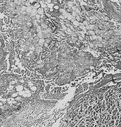
A 45-year-old man developed severe abdominal pain and was transported to the emergency room where he was found to be hypotensive and in shock. He was unresponsive to therapy and died 2 days later. Based upon this history and the accompanying image in below figure, which of the following most likely initiated this sequence of events?
A. acute pancreatitis
B. esophageal varices
C. gastric ulcer
D. liver cirrhosis
E. splenic infarct
Correct Answer: A
Section: Pathology and Path physiology In the lower left corner of figure is pancreas while the rest is composed of necrotic fat cells, some viable fat cells, and inflammatory exudate. Thus, the initiating event in this case is acute pancreatitis. The image does not show any of the other organs listed, thereby ruling out choices B through E.
Question 248:
A 7-year-old girl presents with a neck mass located at the anterolateral aspect of the neck, anterior to the sternocleidomastoid muscle. A biopsy of the lesion revealed a largely cystic mass lined by stratified squamous epithelium surrounded by an underlying dense layer of lymphoid tissue with germinal centers. Based on these findings, what is the most likely diagnosis?
A. branchial cleft cyst
B. granulomatous lymphadenitis
C. Hodgkin lymphoma
D. metastatic laryngeal carcinoma
E. thyroglossal duct cyst
Correct Answer: A
Section: Pathology and Path physiology A cystic structure in the lateral neck lined by squamous or, less usually, columnar epithelium and surrounded by lymphoid issue with germinal centers is invariably a branchial cleft cyst. Granulomatous lymphadenitis (choice B) should demonstrate granulomatous inflammation in a lymph node which is not described here. Hodgkin lymphoma (choice C) occurs in five subtypes, none of which is associated with epithelial tissue. Metastatic laryngeal carcinoma (choice D) is usually squamous cell in origin and would demonstrate pleomorphic polygonal cells with "prickles" and "pearls." A thyroglossal duct cyst (choice E) can have a histological appearance very similar to that of a branchial cleft cyst but the most important difference is the midline location of the thyroglossal duct cyst.
Question 249:
Over the past few months, a 5-year-old boy has developed changes suggestive of puberty. Physical examination reveals the presence of secondary sex characteristics including pubic hair and penile enlargement. Laboratory studies reveal increased levels of ACTH. If you were able to examine his adrenals, which of the following would you most likely find?
A. bilateral atrophy of the cortex
B. bilateral atrophy of the medulla
C. bilateral cortical hyperplasia
D. bilateral medullary hyperplasia
E. nodule in the cortex
F. nodule in the medulla
Correct Answer: C
Section: Pathology and Path physiology This child has one of a group of unusual diseases (congenital adrenal hyperplasia) in which there is an inherited deficiency of an enzyme in the biosynthetic pathway for the corticosteroids; most of these cases are due to a partial deficiency of 21-hydroxylase. The decreased feedback inhibition to the pituitary results in increased production of ACTH, resulting in bilateral adrenal cortical hyperplasia. Impairment of corticosteroid synthesis shunts more substrate into the sex steroid pathway, leading to increased production of androgens (resulting in precocious puberty or virilism in a female child). Bilateral atrophy of the cortex (choice A) has a number of causes. Primary causes are unusual and tend to affect both glucocorticoid and mineralocorticoid production; however, secondary causes (e.g., exogenous steroids) are more common and tend to affect only glucocorticoid production. Precocious puberty is not seen. Bilateral atrophy of the medulla (choice B) with a normal cortex would be extremely unusual and would not be expected to produce precocious puberty. Bilateral medullary hyperplasia (choice D) has been reported but is very unusual and would also not produce precocious puberty. Anodule in the cortex (choice E) could be either a hyperplastic nodule or an adenoma. The former is asymptomatic and the latter is most unlikely to produce precocious puberty but, if it did, would probably also demonstrate hypercortisolism, which is not seen in this patient. A nodule in the medulla (choice F) could be a small, early pheochromocytoma but this would not produce precocious puberty.
Question 250:
A 5-year-old boy is brought to a pediatrician by his mother for progressive abdominal girth and poor appetite. His mother is concerned over a "growing lump" she has felt in his abdomen, and physical examination confirms the presence of a large palpable mass in the left upper quadrant. Abdominal ultrasound demonstrates an 18-cm solid mass in the upper pole of the left kidney. Arenal biopsy is taken. Which of the following is the pathologist most likely to see on microscopic examination of the biopsy?
A. a mixture of proliferating smooth muscle cells, mature fat cells, and abnormalblood vessels
B. numerous fusiform cysts lined by uniform cuboidal cells and no normal parenchyma
C. primitive mesenchymal tissue with minimal tubular and glomerular differentiation
D. sheets of large cells with abundant clear cytoplasm and small uniform nuclei
E. well-formed papillary frond structures lined by cytologically normal, thickened epithelium
Correct Answer: C
Section: Pathology and Path physiology A large tumor appearing in the pole of a kidney in a child of this age is almost certainly a Wilms' tumor. The microscopic appearance of this neoplasm can be highly variable with some areas resembling renal blastema, as well as epithelial and stromal areas. One usually sees attempts to form glomeruli and tubules. Amixture of proliferating smooth muscle cells, mature fat cells, and abnormal blood vessels (choice A) describes an angiomyolipoma. Numerous fusiform cysts lined by uniform cuboidal cells and no normal parenchyma (choice B) is the microscopic appearance of infantile polycystic disease. Sheets of large cells with abundant clear cytoplasm and small uniform nuclei (choice D) describe a renal oncocytoma. Well- formed papillary frond structures lined by cytologically normal, thickened epithelium (choice E) is a description that is compatible with a renal papillary adenoma.
Nowadays, the certification exams become more and more important and required by more and more enterprises when applying for a job. But how to prepare for the exam effectively? How to prepare for the exam in a short time with less efforts? How to get a ideal result and how to find the most reliable resources? Here on Vcedump.com, you will find all the answers. Vcedump.com provide not only USMLE exam questions, answers and explanations but also complete assistance on your exam preparation and certification application. If you are confused on your USMLE-STEP-1 exam preparations and USMLE certification application, do not hesitate to visit our Vcedump.com to find your solutions here.