A 28-year-old male patient suffering from head trauma resulting from a car accident is brought unconscious to the emergency room. In performing the pupillary light reflex, you notice that even though the left pupil constricts when you shine a light directly into the left eye, it does not do so when you shine a light into the right eye. This is best explained by a disconnection between which of the following bilateral structures?
A. Edinger-Westphal nucleus
B. habenula
C. inferior colliculus
D. lateral geniculate nucleus
E. medial geniculate nucleus
Correct Answer: A
Section: Anatomy The central visual pathway for the papillary light reflex is organized as follows: fibers from the ganglionic layer of the retina project posteriorly to the pretectum, which in turn innervates the Edinger- Westphal nucleus. Preganglionic parasympathetic neurons in the Edinger- Westphal nucleus project to the ciliary ganglion, which sends postganglionic parasympathetic innervation back to the constrictor pupillae of the eye. The Edinger-Westphal nuclei from each side of the midbrain are also connected to each other by projections running through the posterior commissure. Disconnection of these fibers will result in loss of the consensual papillary light reflex on the contralateral side, as happens in this case. The habenula (choice B) is a nucleus of the thalamus, which does not participate in the central visual pathways. The lateral geniculate nucleus (choice D) receives fibers from the ganglionic layer of the retina. However, fibers participating in the papillary light reflex run through this structure without synapsing and terminate in the pretectum. Thus, the lateral geniculate nucleus does not participate in the pupillary light reflex. The inferior colliculus (choice C) and the medial geniculate nucleus (choice E) are components of the auditory system.
Question 802:
A premature female infant is born about 24 weeks after fertilization and develops rapid, labored breathing shortly after birth. She is immediately transferred to intensive care where she is diagnosed with hyaline membrane disease (HMD). Which of the following is most likely deficient in the infant?
A. alveolar ducts
B. lung surfactant
C. terminal saccules
D. type I alveolar cells
E. type II alveolar cells
Correct Answer: D
Section: Anatomy HMD is also known as respiratory distress syndrome, which is most often caused by the lack of lung surfactant, due to a premature birth. Lung surfactant production begins around 20 weeks after fertilization. But it is present only in small amounts until the last 2 weeks before birth when its amount increases significantly. Alveolar ducts (choice A) branch from the respiratory bronchioles during development. Type I alveolar cells (choice D) or pneumocytes are squamous epithelial cells, which participate in gas exchange. These epithelial cells line the terminal saccules (choice C). Type II alveolar cells (choice E) synthesize surfactant.
Question 803:
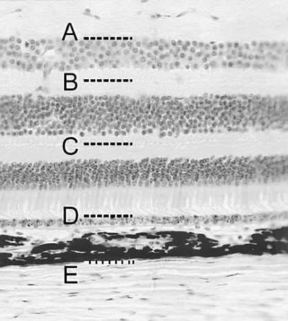
Which label in following figure indicates the typical plane of separation at which retinal detachment occurs?
A. A
B. B
C. C
D. D
E. E
Correct Answer: D
Section: Anatomy Retinal separation typically occurs at the interface between the retinal pigment epithelium and the outer limit of the sensory (neural) retina. The weakness of this plane is attributed to the manner in which the retina develops, a process that involves obliteration of the space between two of the layers of the optic cup--an inner layer from which the sensory retina arises and an outer layer from which the retinal pigment epithelium arises. Other retinal layers are bridged by neuronal processes, and Müller cells (the retina's glial cells) span the entire thickness of the neural retina. Plane A(choice A) marks the boundary between the nerve fiber layer above and the ganglion cell layer below. The nerve fiber layer is composed of axons of the retinal ganglion cells. Plane B (choice B) is within the inner plexiform layer, the site of synaptic contacts between bipolar neurons, retinal ganglion cells, and amacrine cells. Plane C (choice C) is within the outer plexiform layer, the site of synapses among bipolar cells, rods and cones, and horizontal cells. The boundary between the choroid (of the middle vascular tunic or uvea) and the sclera (of the external, fibrous tunic) is marked by plane E (choice E).
Question 804:
During surgery at the root of the neck, an attending surgeon cautions her resident to locate important structures which need to be protected. One of these is the phrenic nerve, responsible for the innervation of the diaphragm and thus, respiration. The phrenic nerve can be positively identified by which of the following anatomical relationships?
A. It is found immediately between the common carotid artery and the internal jugular vein.
B. It lies immediately between the esophagus and the trachea.
C. It lies on the scalenus medius muscle.
D. It wraps around the right subclavian artery.
E. The suprascapular and transverse cervical arteries cross over it anteriorly.
Correct Answer: D
Section: Anatomy At the root of the neck, the phrenic nerve (C3, C4, C5) lies on the scalenus anterior muscle, not the scalenus medius (choice C). The transverse cervical and suprascapular arteries course over it. The vagus (tenth cranial) nerve, not the phrenic nerve is located between the common carotid artery and the internal jugular vein (choice A). The recurrent laryngeal branch from the vagus nerve wraps around the right subclavian artery (choice D), and courses cranially between the esophagus and the trachea (choice B).
Question 805:
Cells in the pancreas that secrete glucagon and insulin are which of the following?
A. A and B cells
B. acinar cells
C. D cells
D. pancreatic D1 cells
E. pancreatic polypeptide cells
Correct Answer: A
Section: Anatomy
In the human pancreas, A and B cells of the islets of Langerhans secrete glucagon and insulin,
respectively. Pancreatic D1 cells (choice D) release a product similar to vasoactive intestinal polypeptide.
Pancreatic polypeptide cells (choice E) secrete pancreatic polypeptide and D cells (choice C) release
somatostatin. All the aforementioned cells belong to the endocrine pancreas. Acinar cells (choice B) are
part of the exocrine pancreas and do not secrete glucagon or insulin.
Question 806:
Below figure is a high magnification photomicrograph of the gall bladder. The arrow points to the internal lining that is formed by which of the following?
A. pseudostratified columnar epithelium
B. simple columnar epithelium
C. stratified cuboidal epithelium
D. stratified squamous epithelium
E. transitional epithelium
Correct Answer: B
Section: Anatomy The lining of the gallbladder is a simple columnar epithelium identifiable by tall cells with elongated nuclei arranged at the same level. Pseudostratified epithelium (choice A) is distinguishable from the simple columnar epithelium by the cell nuclei being arranged at different levels. Stratified cuboidal epithelium (choice C) is characterized by short cells with nuclei arranged at different levels. Stratified epithelium (choice D) has a characteristic cellular basal layer with flat degenerate cells in its upper layer. Transitional epithelium (choice E) is a type of stratified epithelium exclusively confined to the urinary tract.
Question 807:
A 19-year-old man was in a barroom brawl and was punched squarely in the right eye. He comes to the emergency room the next day and complains of diplopia. An X-ray reveals fracture of the orbital floor. Neurological examination shows loss of sensation of the skin of the right face below the right eye and the upper gums. Which of the following nerves may be injured?
A. frontal nerve
B. infraorbital nerve
C. nasociliary nerve
D. supraorbital nerve
E. trochlear nerve
Correct Answer: B
Section: Anatomy The infraorbital nerve, a branch of the maxillary (V2) division of the trigeminal (fifth cranial) nerve, courses below the orbital floor to reach the area of skin below the eye. It provides superior alveolar branches to supply the upper gums and is vulnerable in fractures involving the floor of the orbit and face area. All the nerves mentioned in the other choices will be spared by this type of injury. The frontal nerve (choice A) and nasociliary nerve (choice C) are branches from the ophthalmic division (V1) of the trigeminal (fifth cranial) nerve and course within the orbit. The supraorbital (choice D) nerve is a continuation branch of the frontal nerve onto the forehead, providing sensory innervation for this area. The trochlear (fourth cranial) nerve is also located within the orbit.
Question 808:
Protein zero (P0) is the predominant protein in myelin in the peripheral nervous system and its function is to stabilize adjacent plasma membranes by interaction with similar P0 molecules. Which of the following cells manufacture P0?
A. fibrous astrocytes
B. microglia
C. oligodendrocytes
D. protoplasmic astrocytes
E. Schwann cells
Correct Answer: E
Section: Anatomy Schwann cells produce myelin in the peripheral nervous system whereas oligodendrocytes produce myelin in the central nervous system. Oligodendrocytes manufacture the proteolipid protein, the functional equivalent to P0 in the central nervous system. Fibrous (choice A) and protoplasmic (choice D) astrocytes are supportive cells which play a role in the regulation of brain metabolism. Microglia (choice B) are mesodermal in origin and have phagocytotic activity in the central nervous system.
Question 809:
Aperilunate fracture dislocation is a devastating closed injury of the wrist. It usually results from a fall where the weight of the body is transferred onto the wrist. The hand is caught in the hyperextended and ulnar deviated position. The fracture dislocation involves rupture of interosseous ligaments, joints, and ultimately dislocation/fracture of the lunate bone. In the anatomical position, which carpal bone lies directly distal to the lunate?
A. capitates
B. hamate
C. scaphoid
D. trapezoid
E. triquetrum
Correct Answer: A
Section: Anatomy The capitate bone lies directly distal to the lunate. The mechanism of perilunate fracture dislocation involves rupture of the radioscaphocapitate and scapholunate interosseous ligaments, dislocation of the capitolunate joint, rupture of the lunotriquetral interosseous ligament, and finally dislocation/rupture of the lunate. In the anatomical position, the hamate (choice B) is the most medial carpal bone, located just distal to the triquetrum. The scaphoid (choice C) lies lateral to the lunate and proximal to the trapezium (choice D), the carpal bone articulating with the thumb. The trapezoid (choice E) is medial to the trapezoid and distal to the scaphoid. The lunate bone (choice C) lies adjacent to the scaphoid in the proximal row of carpals and with the scaphoid articulates with the radius at the radiocarpal or wrist joint. It is not related to the anatomic snuffbox. The pisiform bone (choice D) is a sesamoid bone in the tendon of the flexor carpi ulnaris on the lateral wrist. It is not related to the anatomic snuffbox.
Question 810:
A patient was thrown from a tractor, which partially ran over him and caused injury to the base of the skull. The origin of the internal jugular vein at the jugular foramen was compromised. Which of the following cranial nerves courses through the jugular foramen?
A. abducens (sixth cranial) nerve
B. facial (seventh cranial) nerve
C. hypoglossal (twelfth cranial) nerve
D. spinal accessory nerve (eleventh cranial) nerve
E. vestibulocochlear (eighth cranial) nerve
Correct Answer: D
Section: Anatomy The spinal accessory (eleventh cranial) nerve takes its origins in the neck, but then runs cranially into the skull through the foramen magnum to join with its cranial component. They exit as one through the jugular foramen, along with the glossopharyngeal (ninth cranial) and vagus (tenth cranial) nerves. The abducens (sixth cranial) nerve (choice A) runs through the superior orbital fissure to reach the orbit. The facial (seventh cranial; choice B) and vestibulocochlear (eighth cranial; choice E) nerves run together through the internal acoustic meatus into the temporal bone. The facial nerve exits the skull through the stylomastoid foramen. The hypoglossal (twelfth cranial; choice C) nerve exits the skull through the hypoglossal canal.
Nowadays, the certification exams become more and more important and required by more and more enterprises when applying for a job. But how to prepare for the exam effectively? How to prepare for the exam in a short time with less efforts? How to get a ideal result and how to find the most reliable resources? Here on Vcedump.com, you will find all the answers. Vcedump.com provide not only USMLE exam questions, answers and explanations but also complete assistance on your exam preparation and certification application. If you are confused on your USMLE-STEP-1 exam preparations and USMLE certification application, do not hesitate to visit our Vcedump.com to find your solutions here.