In the skull, a network of thick-walled vessels named dural venous sinuses drains the cerebrospinal fluid and the venous blood from the brain. These vessels are formed by reflections of the dura mater, which also form partitions between major parts of the brain. Which of the following dural venous sinuses is associated with the falx cerebri?
A. cavernous sinus
B. inferior petrosal sinus
C. sigmoid sinus
D. superior sagittal sinus
E. transverse sinus
Correct Answer: D
Section: Anatomy
The falx cerebri is the dural partition separating the hemispheres of the brain, above the corpus callosum.
It has in its superior edge the superior sagittal sinus and in its inferior border, the inferior sagittal sinus. The cavernous sinus (choice A) is a dural pocket in the middle cranial fossa and is located on either side of the sella turcica. It is unusual because it contains the internal carotid artery and cranial nerves III, IV, V1, V2, and VI. The sigmoid sinus (choice C) is located in the posterior cranial fossa, below the level of the tentorium cerebelli, which contains in its posterior border the transverse sinus (choice E).
Question 812:
The most common type of testicular cancer is germ cell carcinoma. However, testicular tumors arising from other cell types in the testis also occur. Arrow 1 in following figure, indicates which of the following structures?
A. Leydig cell
B. Sertoli cell
C. spermatid
D. spermatocyte
E. spermatogonia
Correct Answer: A
Section: Anatomy Arrow 1 indicates an interstitial cell of Leydig. These cells are recognizable by their location in the intertubular space, close to the capillaries and lymph vessels. Sertoli cells (choice B) are columnar cells with a large nucleolus. They span the distance from the seminiferous tubular wall to the lumen. However, their nuclei are located closer to the tubular wall. Spermatids (choice C; arrow 4) have a condensed nucleus, which becomes elongated with further maturation. This cell type is located closest to the lumen of the seminiferous tubule. Spermatocytes (choice D; arrow 2) are seen above the layer spermatogonia (choice E; arrow 3) which are located against the basal lamina.
Question 813:
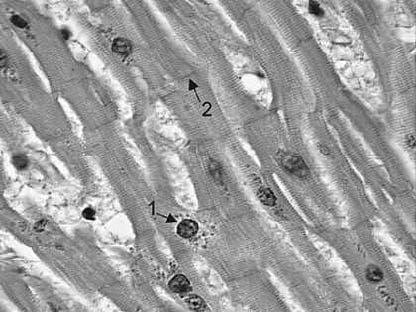
Referring to following figure, arrow 2 indicates which of the following structures?
A. intercalated disk
B. motor end-plate
C. sarcoplasmic reticulum
D. tendinous junction
E. transverse tubule or T tubule
Correct Answer: A
Section: Anatomy The intercalated disks are specialized junctional complexes found only in cardiac muscle and they appear as dark lines between the muscle fibers. The motor end-plate (choice B) is a specialized group of synapses between the axon terminals of a motor neuron and the sarcolemma of a skeletal muscle fiber. It is not seen in cardiac muscle. The sarcoplasmic reticulum (choice C) is a specialized modification of the smooth endoplasmic reticulum for sequestering calcium ions. The transverse tubule or T tubule (choice E) is an invagination of the sarcolemma, which penetrates the muscle fiber and overlies the surface of the myofibrils. The sarcoplasmic reticulum and T tubule can only be seen in electron micrographs. There is no tendinous junction (choice D) in cardiac muscle.
Question 814:
In emphysema, which of the following components of the bronchioles is affected?
A. ciliated cuboidal epithelial cells
B. Clara cells
C. elastic fibers
D. goblet cells
E. squamous type I alveolar epithelial cells
Correct Answer: C
Section: Anatomy Elastic fibers are destroyed in emphysema by elastase. This protease is released by neutrophils recruited by macrophages under abnormal stimulus such as cigarette smoke. The loss of elasticity in the bronchioles and alveolar walls gives rise to emphysema, characterized by chronic airway obstruction. Ciliated cuboidal epithelial (choice A) and Clara (choice B) cells line the terminal bronchioles. Goblet cells (choice D) may be found at the beginning of the bronchioles and squamous type I alveolar epithelial cells line the respiratory bronchioles.
Question 815:
During development, the notochord grows in a cranial direction until it reaches the prechordal plate. This plate is the primordium of the oropharyngeal (or buccopharyngeal) membrane, which, in the embryo, will separate the stomodeum from the foregut. At 26 days of gestation, the oropharyngeal membrane will break down, allowing communication of the foregut with the oral cavity. Of the following structures in the adult, which one lies at the same location as the embryonic oropharyngeal membrane?
A. buccinators
B. palatoglossus
C. palatopharyngeus
D. stylopharyngeus
E. superior constrictor
Correct Answer: B
Section: Anatomy The palatoglossus muscle, which can be observed in the oral cavity to form the palatoglossal arch anterior to the palatine tonsil, lies in the same location as the embryonic oropharyngeal membrane. It lies at the junction line between the stomodeum and the foregut. The buccinator (choice A) is a muscle of the cheek and thus is located in the original stomodeum. The palatopharyngeus (choice C) is located posterior to the palatoglossus and palatine tonsil, forming the palatopharyngeal arch. The palatopharyngeus, stylopharyngeus (choice D), and superior constrictor (choice E) muscles are all pharyngeal muscles and thus are located in the original foregut.
Question 816:
Which of the following is the correct sequence of erythroid differentiation?
A. proerythroblast, basophilic erythroblast, polychromatophilic erythroblast, normoblast, reticulocyte, mature erythrocyte
B. proerythroblast, normoblast, reticulocyte, polychromatophilic erythroblast, basophilic erythroblast, mature erythrocyte
C. proerythroblast, polychromatophilic erythroblast, basophilic erythroblast, reticulocyte, normoblast, mature erythrocyte
D. proerythroblast, reticulocyte, normoblast, polychromatophilic erythroblast, basophilic erythroblast, mature erythrocyte
E. proerythroblast, reticulocyte, polychromatophilic erythroblast, normoblast, basophilic erythroblast, mature erythrocyte
Correct Answer: A
Section: Anatomy The correct sequence of erythroid differentiation is indicated by choice A. Erythrocyte differentiation in the adult occurs exclusively in the bone marrow and consists of several cellular changes. The cell size decreases: proerythroblast 1419 m in diameter; basophilic erythroblast 1316 m; polychromatophilic erythroblast 1215 m; normoblast, reticulocyte, and mature erythrocyte 810 m. Condensation of the nuclear chromatin and decrease in nuclear diameter occur from the proerythroblast to the normoblast stage with ejection of the nucleus. Subsequent ejection of remaining organelles occurs in the reticulocyte to give rise to the mature erythrocyte. The maturing cells change their staining affinity because the increased hemoglobin in the cytoplasm results in increased acidophilia, whereas the decrease in the ribosome numbers in the cytoplasm results in decreased basophilia. Choices B, C, D, and E are incorrect sequences.
Question 817:
An elderly resident of a nursing home fell down the front steps and subsequently became disoriented and lethargic. He is brought to the emergency room where an emergency MRI reveals that he has developed hydrocephalus due to a small hemorrhage obstructing the foramina of Monro. The foramina of Monro allow for communication between which of the following?
A. fourth ventricle and cerebral aqueduct
B. fourth ventricle and subarachnoid space
C. lateral ventricles and third ventricle
D. third ventricle and cerebral aqueduct
E. third ventricle and fourth ventricle
Correct Answer: C
Section: Anatomy The foramina of Monro form the communication between the lateral ventricles and the third ventricle. The cerebral aqueduct of Sylvius flows caudally into the fourth ventricle (choice A). The lateral foramina of Luschka and the median foramen of Magendie allow for communication between the fourth ventricle and the subarachnoid space. The third ventricle communicates posteriorly with the cerebral aqueduct of Sylvius (choice D). Thus, the third and fourth ventricle communicate by way of this cerebral aqueduct.
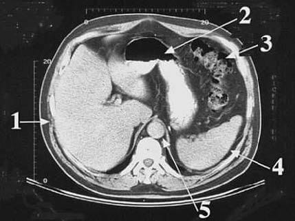
Question 818:
Arrow 4 in following figure, is pointing to which of the following structures?
A. abdominal aorta
B. colon
C. liver
D. spleen
E. stomach
Correct Answer: D
Section: Anatomy The spleen (arrow 4) lies to the left of the abdominal cavity. It is in contact with the left side of the stomach (arrow 2) and lodges against the left paravertebral gutter. The abdominal aorta (choice A, arrow 5) is seen as the circular structure immediately anterior to the vertebra. The colon (choice B, arrow 3) is the convoluted structure to the left anterior aspect of the abdominal cavity. The large liver (choice C, arrow 1) occupies most of the right side of the abdominal cavity. The stomach (choice E, arrow 2) is located between the colon and the liver, and in this case, contains liquid contrast material.
Question 819:
During a direct inguinal hernia repair operation, the attending surgeon reminds the firstyear surgical resident that an anatomical variation for the origin of the obturator artery exists. This artery normally arises from the internal iliac artery but it may also originate directly from which of the following vessels?
A. common iliac artery
B. external iliac artery
C. inferior epigastric artery
D. inferior vesical artery
E. superior vesical artery
Correct Answer: C
Section: Anatomy The obturator artery can originate from the inferior epigastric artery, a location which renders it vulnerable during inguinal hernia surgical repair. The common iliac artery (choice A) only has two branches, the external and internal iliac arteries. Within the pelvis, the external iliac artery (choice B) gives out two branches, the deep circumflex iliac and inferior epigastric arteries. The superior (choice E) and inferior (choice D) vesical arteries are branches of the internal iliac arteries, supplying the bladder.
Question 820:
A 37-year-old rural female patient developed pain in the lower abdomen and pelvic regions. Her physician suspects a ruptured ectopic pregnancy. However, because of the isolation of the rural community, no medical imaging or laboratory procedure is available and the physician decides to perform a culdocentesis. In the latter procedure, the needle will aspirate from which of the following spaces?
A. ovarian fossa
B. rectouterine pouch
C. uterine body
D. uterine cervix
E. vesicouterine pouchAnswer:
Correct Answer: B
Section: Anatomy In culdocentesis, the needle is inserted through the posterior fornix of the vagina and fluid is aspirated from the rectouterine pouch. If nonclotting blood is collected then the likelihood of a ruptured ectopic pregnancy is high. This procedure is rapid and inexpensive, however, serum progesterone level assay or ultrasonography are preferred methods. The ovarian fossa (choice A) or vesicouterine pouch (choice E) are not used in culdocentesis. The uterine body (choice C) and cervix (choice D) would not reveal blood from a ruptured ectopic pregnancy and thus are also not used in culdocentesis.
Nowadays, the certification exams become more and more important and required by more and more enterprises when applying for a job. But how to prepare for the exam effectively? How to prepare for the exam in a short time with less efforts? How to get a ideal result and how to find the most reliable resources? Here on Vcedump.com, you will find all the answers. Vcedump.com provide not only USMLE exam questions, answers and explanations but also complete assistance on your exam preparation and certification application. If you are confused on your USMLE-STEP-1 exam preparations and USMLE certification application, do not hesitate to visit our Vcedump.com to find your solutions here.