A 48-year-old female patient is brought to the emergency room by her husband. He reports that his wife suffers from hypertension but, as a high-level executive with a lot of pressure at work, she has been neglecting to take her medication. This morning, as he entered the garage to leave for work, he found his wife lying on the ground next to her own car. She was experiencing uncontrolled flailing of the left arm and leg. What is the most likely site of brain lesion in this patient?
A. anterior limb of the left internal capsule
B. anterior limb of the right internal capsule
C. cerebellum
D. left subthalamic nucleus of Luys
E. right subthalamic nucleus of Luys
Correct Answer: E
Section: Anatomy Hemiballismus is a movement disorder characterized by involuntary large amplitude movements of one or both limbs on one side of the body. It results from infarct damage to the contralateral subthalamic nucleus of Luys, in this case the right one. The left subthalamic nucleus (choice D) controls the limbs on the right side of the body, which are not affected in this case. The anterior limbs of the internal capsule (choices Aand B) contain mainly thalamocortical and corticothalamic fibers and lesions in these areas do not result in hemiballismus. Lesions in the cerebellum (choice C) also do not result in hemiballismus.
Question 822:
In cleaning the teeth in a patient, a dental hygienist accidentally cuts the gums of the posterior two molar teeth in the lower jaw on the lateral side. The pain of this injury is registered by which of the following nerves?
A. anterior, middle, and posterior superior alveolar nerves
B. buccal nerve
C. greater palatine nerve
D. lingual nerve
E. nasopalatine nerve
Correct Answer: B
Section: Anatomy The gums on the lateral side of the mandibular molar teeth are innervated by the buccal nerve (long buccal nerve). All three superior alveolar nerves (choice A) supply the gums lateral to all maxillary teeth. The greater palatine nerve (choice C) innervates the gums medial to the maxillary premolar and molar teeth. The lingual nerve (choice D) supplies the gums medial to all mandibular teeth. The nasopalatine nerve (choice E) innervates the gums posterior to the maxillary incisors.
Question 823:
Recanalization of the bile duct after the 13th week after fertilization allows for bile produced in the liver to reach the duodenum. However, if recanalization fails to occur and this cannot be corrected surgically, the affected infant will need a liver transplant. During development, the liver arises from which of the following?
A. foregut
B. hindgut
C. midgut
D. pleuroperitoneal membrane
E. septum transversum
Correct Answer: A
Section: Anatomy The liver arises as a ventral outgrowth from the caudal portion of the foregut. The midgut (choice C) arises past the junction point between the bile duct and the duodenum, distal to the formative outgrowth of the liver. The midgut gives rise to the small intestine and part of the large intestine. The hindgut (choice B) arises further distally and gives rise to the rest of the large intestine, the superior part of the anal canal, the epithelium of the urinary bladder, and most of the urethra. The pleuroperitoneal membrane (choice D) and the septum transversum (choice E) are developmental components of the diaphragm.
Question 824:
A professional football player was diving for a touchdown when his face mask was grabbed and wrenched, causing neck hyperextension and rotation to the right. When brought to the sideline, the player complained of a burning sensation radiating down the right upper extremity and neurological examination revealed right lateral weakness of this limb. Movements affected were arm rotation and flexion, elbow flexion, forearm supination, and thumb flexion. The patient is diagnosed with a brachial plexus injury at the level of C6. 29. Which of the following muscles can perform arm and elbow flexion along with forearm supination?
A. biceps brachii
B. brachialis
C. brachioradialis
D. coracobrachialis
E. supinator
Correct Answer: A
Section: Anatomy The biceps brachii muscle attaches proximally by its short head to the coracoid process of the scapula and by its long head to the supraglenoid tubercle. Distally it attaches by a strong tendon to the tuberosity of the radius and by an aponeurosis to the ulna. It thus can perform arm and elbow flexion along with forearm supination. The brachialis (choice B) attaches proximally to the anterior aspect of the lower half of the humerus and distally to the coronoid process of the ulna. It can only perform elbow flexion. The brachioradialis (choice C) attaches from the lateral supracondylar ridge of the humerus to the base of the styloid process of the radius. Although innervated by the nerve to the extensor compartment, the radial nerve, it performs elbow flexion and forearm pronation. The coracobrachialis (choice D) attaches from the coracoid process of the scapula to the anterior aspect of the upper half of the humerus. It performs arm flexion and weak adduction. The supinator (choice E) attaches proximally to the lateral epicondyle of the humerus and the annular ligament of the radius. Distally, it covers nearly the upper third of the radius and attaches to its lateral anterior aspect. It supinates the forearm, but is a weaker supinator than the biceps brachii.
Question 825:
An infant is born anencephalic. He presents without both a forebrain and a cerebrum. The remaining brain tissue is exposed, not covered by bone or skin. The infant is blind, deaf, unconscious, and unable to feel pain. Because the infant has a rudimentary brainstem, reflex actions such as respiration (breathing) and responses to sound or touch occur. However, the lack of a functioning cerebrum permanently rules out the possibility of ever gaining consciousness. Anencephaly is the result of a defect in which of the following?
A. closure of the caudal neuropore
B. closure of the rostral neuropore
C. formation of the first branchial arch
D. formation of the somites
E. fusion of the metopon
Correct Answer: B
Section: Anatomy Malclosure of the rostral neuropore during the fourth week of development results in anencephaly and is lethal in the affected newborn. The condition is better termed meroanencephaly because of the presence of the rudimentary brainstem with some functioning nervous tissues. Defects in the closure of the caudal neuropore (choice A) result in varying conditions of spina bifida at the lower end of the spinal cord. The first branchial arch (choice C) and the somites (choice D) do not play any role in the formation of the brain. Fusion of the metopon or forehead (choice E) occurs after birth and also does not play a role in brain formation.
Question 826:
As the consulting physician to the US Open, you are asked to examine a golfer who complains of increased pain with right wrist flexion and pronation activities. The patient also reports discomfort even when simply shaking hands with someone. Examination reveals also decreased sensation in the territory of the ulnar nerve. Your diagnosis is golfer's elbow, affecting mostly the superficial flexor muscles of the forearm. This group of muscles has a common origin from which of the following bony landmarks?
A. head of the radius
B. lateral epicondyle of the humerus
C. medial epicondyle of the humerus
D. olecranon process of the ulna
E. tuberosity of the radius
Correct Answer: C
Section: Anatomy The superficial layer of flexor muscles of the forearm all originate from the medial epicondyle of the humerus. Thus, this condition is also called medial epicondylitis and the most common finding is tenderness with palpation over the anterior aspect of the medial epicondyle. The muscles involved are most often the Pronator Teres, Flexor Carpi Radialis, and Palmaris Longus. The Flexor Digitorum Superficialis and Flexor Carpi Ulnaris may also be affected. There is no muscle attachment to the head of the radius (choice A). The lateral epicondyle of the humerus (choice B) is the attachment point of the common extensor tendon. The olecranon process of the ulna (choice D) is the attachment point for the Triceps Brachii, Flexor Carpi Ulnaris, and Anconeus. The tuberosity of the radius (choice E) receives the distal tendon of the biceps brachii.
Question 827:
A stroke resulting from obstruction of the structure indicated by arrow 1 in following figure, may result in ischemia in which of the following brain regions?
A. Broca's area in the left frontal lobe
B. cerebellum
C. medial aspect of the right frontal lobe
D. pons
E. Wernicke's area in the left frontal lobe
Correct Answer: C
Section: Anatomy Arrow 1 points to the right internal carotid artery which supplies the anterior and middle cerebral arteries in the brain. The territory of the right anterior cerebral artery includes the rightmedial aspect of the frontal lobe, which will be affected by obstruction of the internal carotid artery. Broca's (choice A) and Wernicke's (choice E) areas are located in the majority of the population in the left cerebral hemisphere and are supplied by the left middle cerebral artery from the left internal carotid artery. They will not be affected in this case. The cerebellum (choice B) and pons (choice D) receive their blood supply from the basilar artery (arrow 2) which is formed from the vertebral arteries.
Question 828:
In the brain, the amygdala plays an important role in emotional processing. Patients with lesion of the amygdala display impairment in enhanced perception of emotionally salient events. Which of the following is a major output pathway from the amygdala?
A. fasciculus arcuatus
B. fasciculus cuneatus
C. fasciculus of Vicq d'Azyr
D. fornix
E. stria terminalis
Correct Answer: E
Section: Anatomy The stria terminalis or fasciculus of Foville is one of the major output pathways from the amygdala to the septal, hypothalamic, and thalamic nuclei. Another main amygdaloid output pathway is the ventral amygdalofugal pathway. The fasciculus arcuatus (choice A) or superior longitudinal fasciculus is a bundle of fibers in the cerebrum connecting ipsilateral regions of the frontal, temporal, parietal, and occipital lobes. The fasciculus cuneatus (choice B) carries ascending sensory fibers in the dorsal funiculus of the spinal cord and terminates in the nucleus cuneatus of the medulla oblongata. The fasciculus of Vicq d'Azyr (choice C) or mammillothalamic tract connects the mammillary bodies to the anterior nuclei of the thalamus. This bundle of fibers forms part of Papez circuit, which is also involved in emotional processing. Another part of Papez circuit is the fornix (choice D), a large efferent pathway from the hippocampus.
Question 829:
A 48-year-old male patients is brought to the emergency room because of intense pain of the right face and neck with transient visual loss of the right eye. On examination, the patient has palsy of the oculomotor nerve on the right side with resulting diplopia, along with a right lateralized painful Horner syndrome. This constellation of signs is suggestive of a cervical carotid dissection, which is a separation of the arterial tunical intima from the subjacent tunica media. Which numbered structure in following figure, is the tunica intima?
A. 1
B. 2
C. 3
D. 4
E. 5
Correct Answer: D
Section: Anatomy Arrow 3 points to the tunica intima. The carotid artery is an elastic artery, which contains the following layers aside from the tunica intima: Tunica externa (arrow 1), and tunica media (arrow 5). In a carotid dissection, the tunica intima can elevate or separate from the tunica media with accompanying hemorrhage of the arterial wall. The most common clinical signs are ophthalmological manifestations including painful Horner syndrome, palsy of the oculomotor nerve, diplopia, and transient monocular visual loss. Arrow 2 points to a vasa vasorum, vessels which nourish the thick wall of the aorta. Arrow 4 points to the adipose tissue in the tunica externa.
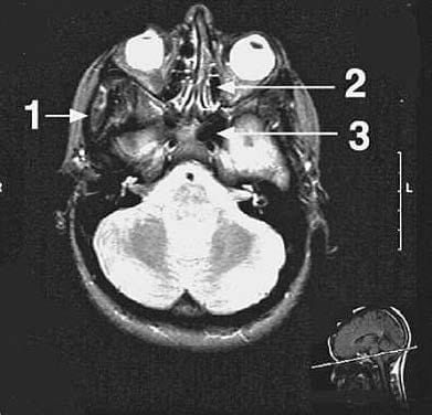
Question 830:
The structure indicated by arrow 2 in following figure, is which of the following?
A. ethmoidal sinus
B. inferior nasal meatus
C. infratemporal fossa
D. maxillary sinus
E. sphenoidal sinus
Correct Answer: A
Section: Anatomy This axial scan is at the level of the orbits as indicated by the insert at the bottom right and the eyeballs in the orbits. Arrow 2 points to the ethmoidal sinus located medial to the orbits. The sinus is divided into compartments by the air cells. The maxillary sinus (choice D) and the inferior nasal meatus (choice B) are located inferior to the level of this scan and are not seen. The sphenoidal sinus (choice E) is indicated by arrow 3 and the infratemporal fossa (choice C) by arrow 1.
Nowadays, the certification exams become more and more important and required by more and more enterprises when applying for a job. But how to prepare for the exam effectively? How to prepare for the exam in a short time with less efforts? How to get a ideal result and how to find the most reliable resources? Here on Vcedump.com, you will find all the answers. Vcedump.com provide not only USMLE exam questions, answers and explanations but also complete assistance on your exam preparation and certification application. If you are confused on your USMLE-STEP-1 exam preparations and USMLE certification application, do not hesitate to visit our Vcedump.com to find your solutions here.